Task: Implementing a comprehensive treatment plan and effective interaction between the clinician and the patient.
Problem: When discussing the clinical situation, the orthodontist uses plaster models of the jaws and which is often complicated for the patient to understand for the patient.
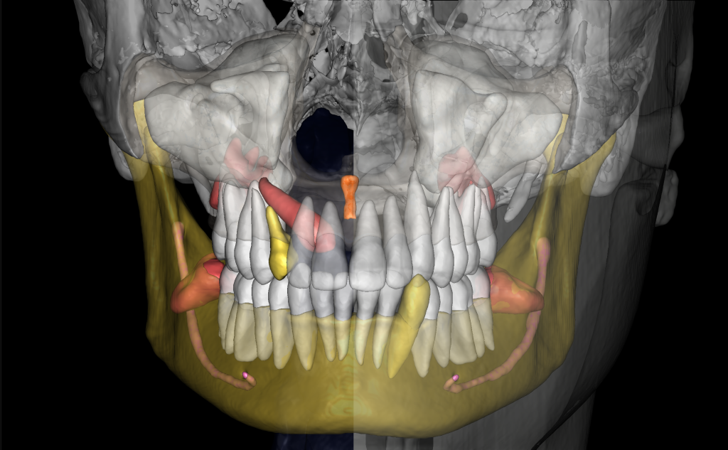
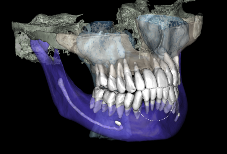
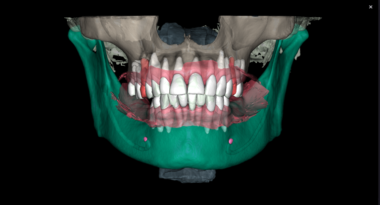
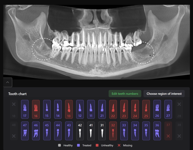
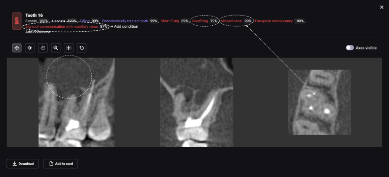
Solution: As an alternative to traditional jaw models, Diagnocat suggests using 3D segmentation, generated based on cone-beam computed tomography (CBCT). In doing so, the patient can see their own face, teeth and bone. The viewing program reduces the transparency of the soft tissue and shows the patient the pathology clearly. A visual 3D model simplifies communication, helps the patient understand the problem, and helps to set the patient up for timely treatment. “
