Task: Establish communication with the patient and plan the autotransplantation of the third molar into the position of tooth 37 (Universal 18)
Problem: The patient has doubts about the necessity of tooth extraction
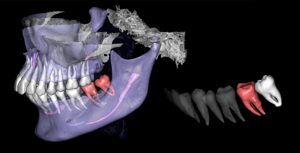
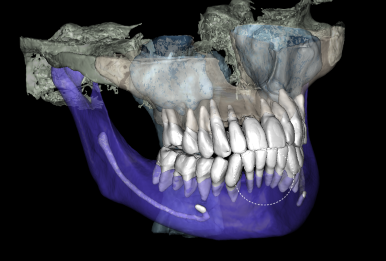
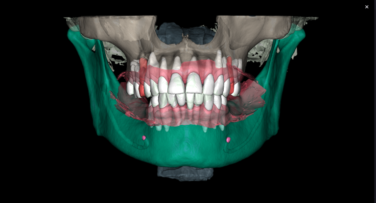
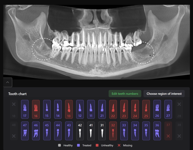
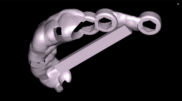
Solution: The automated process of segmentation and formation of 3D models from DICOM files allows extracting individual structures for subsequent 3D printing. The printed model of the third molar, taken from the “STL” module of Diagnocat, is used to prepare the socket for the transplanted tooth. The 3D reconstruction generated using Diagnocat displays the structure of the jaws and teeth and enables the visualization of tooth 37 (Universal 18) with periapical lesion around the roots. In this case, Diagnocat serves as a communication tool that helps convince the patient of the importance of timely implementation of the proposed treatment plan.
