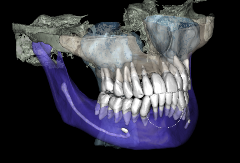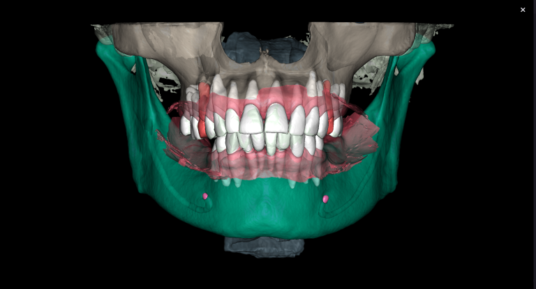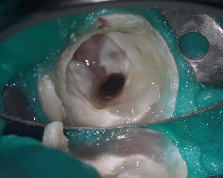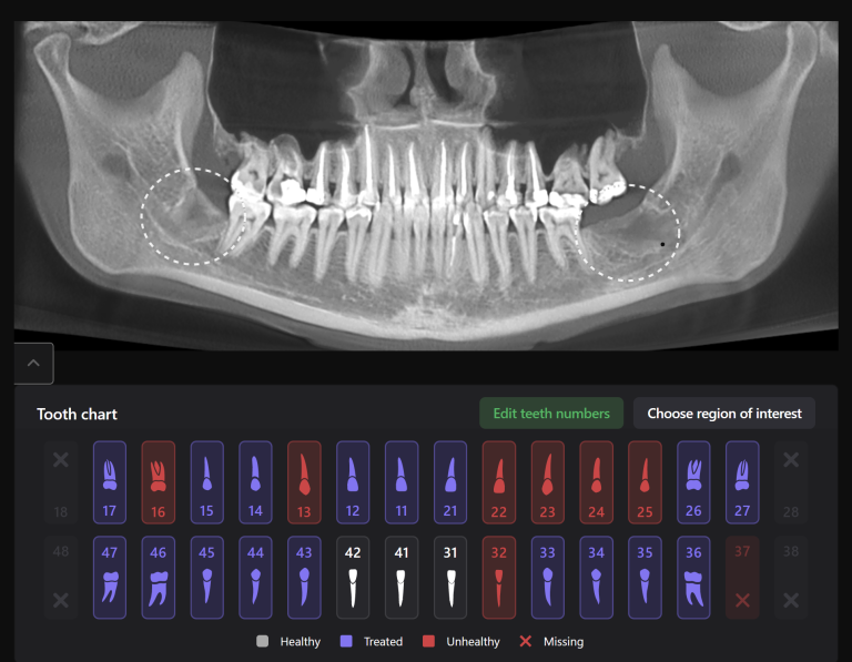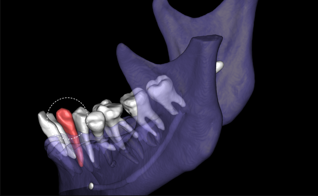
X-ray artifacts should not interfere with precise diagnosis! The 3D reconstruction created by Diagnocat shows tooth 43 (Universal 27), which will be discussed in this clinical case
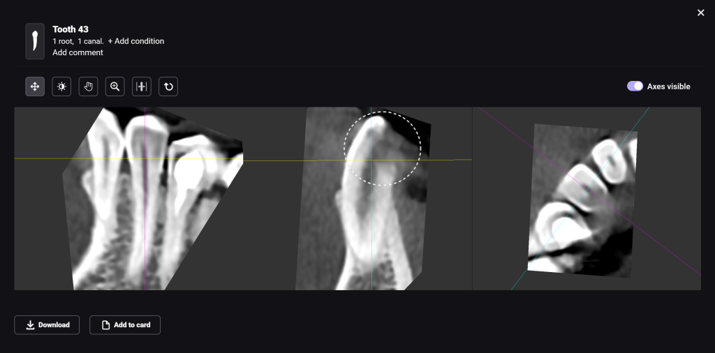
A CBCT scan was performed as part of the checkup, and a radiolucent area can be seen in the coronal part of tooth 43 (Universal 27)
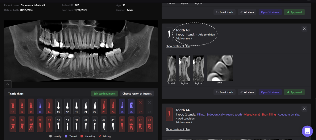
No pathology was found in the area of tooth 43 (Universal 27), since Diagnocat accurately differentiated the artifact from the true pathology
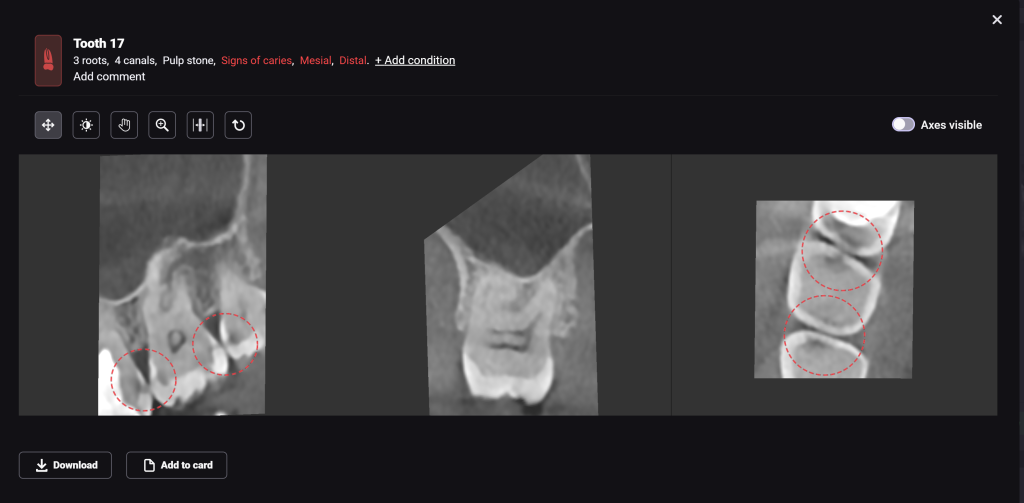
Alternatively, let’s take a look at the tooth 17 (Universal 2) using 3D-Viewer. We can see radiolucency of enamel and dentin, which was correctly indicated as caries in the report. 3D-Viewer is a tool with an interactive coordinate system that allows you to visualize the area of interest simultaneously from three planes: axial, bucco-lingual and mesio-distal.


