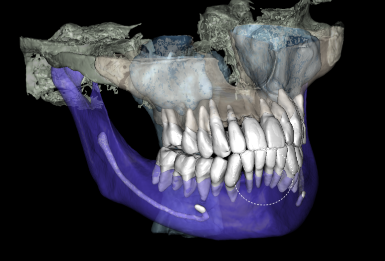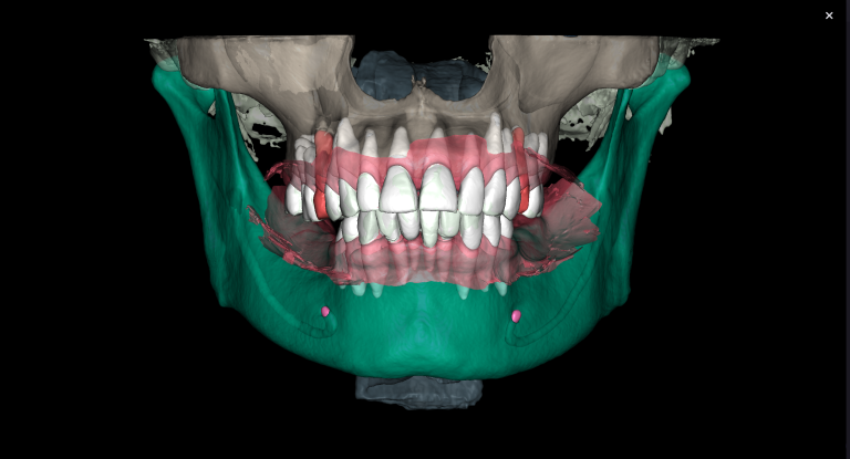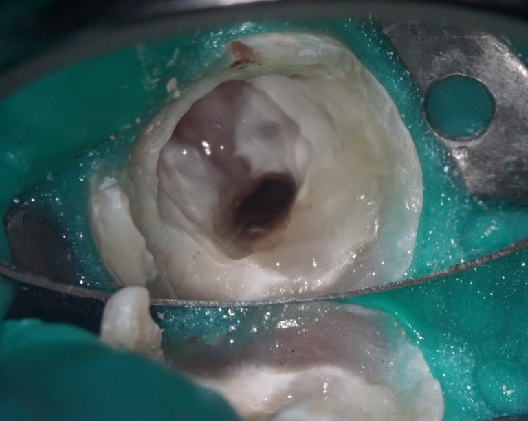
Why do we use CBCT? Here is a clinical case report of a patient who came by for a routine checkup.
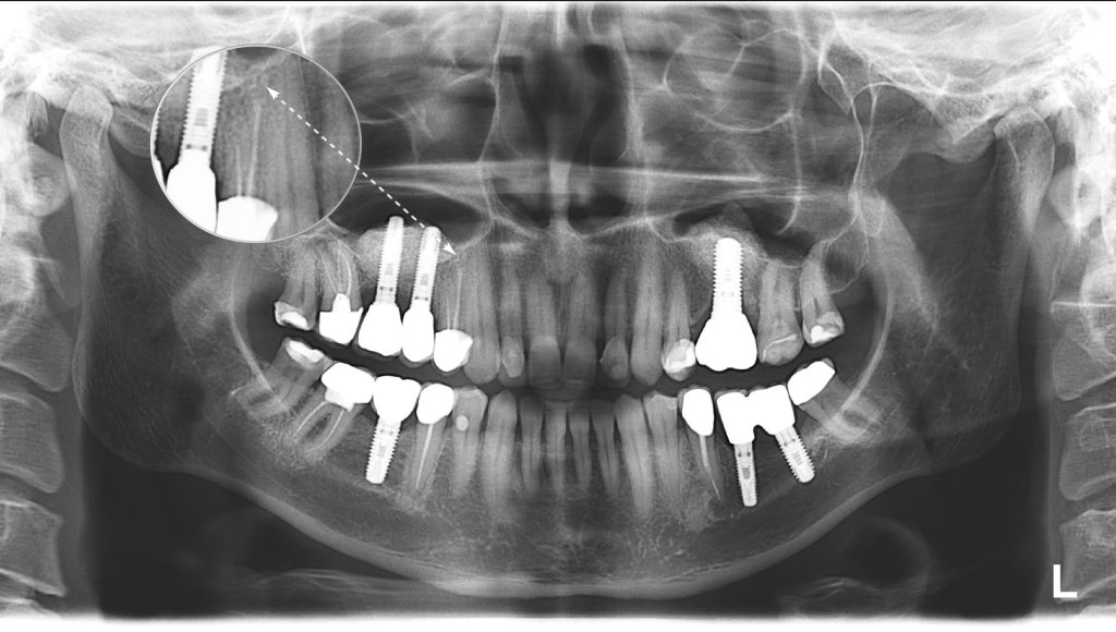
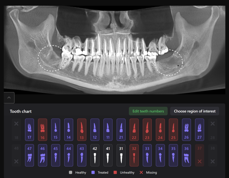
A panoramic dental X-Ray was performed to determine the appropriate treatment. No visible pathology is evident near tooth 14 (Universal 5).

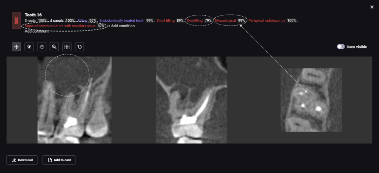
A CBCT scan was performed as part of the checkup, and a radiological screening was created using Diagnocat AI. The report revealed a periapical lesion, which cannot be seen in an ordinary panoramic X-ray.
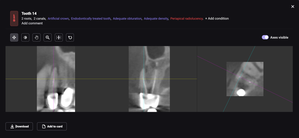
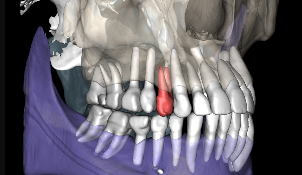
A 3D-Viewer allows the clinician to evaluate the radiological findings interactively and align the axes according to the area of interest.

