A Second Set of Eyes: Using Diagnocat's FDA-Cleared CBCT Tools in Endodontic Practice
Clinical Perspective
Dr. Jayati Pandey, BDS, MDS1
1 Endodontist, Berkeley, California, USA
A 52-year-old patient presents with intermittent dull pain in the lower right quadrant. Tooth #30 has a history of endodontic treatment completed three years ago. The periapical radiograph is equivocal: there is a faint radiolucency at the furcal area, but it is difficult to determine whether this represents healing granulation tissue, a persistent lesion, or simply anatomical noise. The patient is anxious, skeptical of retreatment, and not convinced there is actually a problem.
Most endodontists and general practitioners will recognize this scenario immediately. The clinical suspicion is there, but the two-dimensional image does not provide the clarity needed to make a confident recommendation, let alone explain it to the patient in a way that motivates action. A CBCT is the logical next step. But even with three-dimensional data in hand, two challenges remain: interpreting hundreds of slices efficiently and translating what you find into something the patient can see, trust and understand.
Periapical radiolucency, in particular, can be subtle in its early stages or masked by overlapping anatomy in certain planes. The volume of data in a single CBCT scan is enormous, and the reality of daily practice is that fatigue, time pressure, and case volume introduce inconsistencies that even experienced clinicians acknowledge. This is where AI-supported tools start to make sense: not intended as replacements for clinical judgment, but as a structured second set of eyes.

From Upload to Insight
Diagnocat generates a structured radiological report: a panoramic reconstruction, a color-coded tooth chart, and automatic multiplanar reconstructions for every tooth across axial, vestibulo-oral, and mesio-distal planes within minutes of uploading the DICOM file. The platform is compatible with CBCT systems from Sirona, Vatech, Planmeca, Carestream, and Instrumentarium, among others, so there is no additional hardware to purchase.
Returning to our patient with tooth #30: The first thing I see is the color-coded dental formula. Tooth #30 is flagged red, meaning the software has highlighted it as a region of interest that is suspicious for periapical radiolucency. At a glance, I have a full-mouth overview that directs my attention precisely where it needs to go, rather than requiring me to scroll through every slice hoping I do not miss something at the end of a long day.

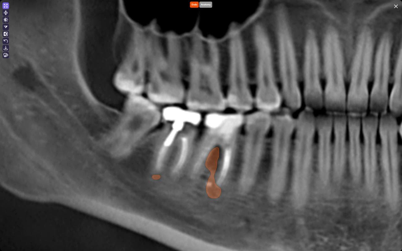
Clicking on tooth #30 opens its detailed report card. Diagnocat's Endo Critical Area Tags highlight the area of suspected periapical radiolucency directly on the tooth slices. The software is not diagnosing; it is highlighting a region that warrants closer clinical evaluation. But that visual flag gives me a clear starting point instead of a manual hunt.

The automatically generated multiplanar reconstructions eliminate what used to be one of the most time-consuming parts of CBCT review: manually setting up cross-sections for each tooth of interest. Diagnocat produces a comprehensive set of slices in three planes for every tooth, with no manual setup required. For tooth #30, I can immediately examine the periapical region from multiple angles, assess the extent of the radiolucency, and evaluate proximity to the anatomical landmarks without toggling between software tools or rebuilding views from scratch.
The built-in MPR tools let me rotate the tooth along all axes, adjust slice orientation with precision, and generate additional custom slices if I need a closer look at a specific area. For complex cases involving root resorption, missed canals, or proximity to the mandibular canal, this level of integrated spatial analysis keeps everything in one place.
The 3D Viewer takes it further. It automatically segments teeth, pulp, and anatomical structures of the maxillofacial region, producing interactive, color-coded models. I can isolate individual teeth, toggle anatomical layers, and view the full craniomaxillofacial anatomy in three dimensions. The platform also generates exportable STL files for educational purposes only, which are useful for team discussions and patient communication, though not intended for clinical treatment planning.
Show and Tell
Back to our patient: she is sitting in the chair, arms crossed, unconvinced. I have seen the periapical radiolucency clearly on the CBCT, with AI supporting my clinical assessment, and I am confident retreatment is indicated. But she is staring at a grayscale image that means nothing to her.
With Diagnocat, I turn the screen toward her and show her the panoramic view with the colored overlay marking the area of concern around tooth #30. Suddenly, the abstract becomes concrete. She can see the highlighted region. She can point to it. I switch to the 3D model, rotate the anatomy, and show her exactly where the lesion sits in relation to her tooth root and the surrounding bone. The structured, print-ready report gives her something to take home, review with her family, and reference when she calls back to schedule.
In my experience, this shift in patient understanding is not marginal. When patients see their condition clearly, they stop feeling like they are being sold a procedure and start making informed decisions about their own health. Anxiety decreases. Questions become more specific and productive. Treatment acceptance improves. This is what happens when you remove the communication barrier between clinician knowledge and patient comprehension.
Consistency Where It Counts
Beyond individual cases, there is a broader practice-level benefit worth acknowledging. AI does not get fatigued at 4 PM on a Friday. It does not rush the last scan before lunch. It applies the same analytical rigor to every tooth in every scan, every time. For practices processing multiple CBCTs daily, that consistency is a genuine safeguard against the natural variability of human attention.
Diagnocat is also HIPAA and SOC 2 compliant, with end-to-end encryption and healthcare-grade cloud infrastructure. The platform runs entirely in the browser, requires no specialized local software, and the latest version integrates with existing practice management systems through the Diagnocat Panel. For U.S. practices navigating increasingly strict data security requirements, that regulatory alignment and ease of deployment matter.
Diagnocat does not replace clinical judgment. It supports it. The FDA-cleared Radiological Report and 3D Viewer give clinicians a faster, more consistent, and more visual way to work through CBCT data, and that clarity extends beyond the clinician's screen to the patient sitting in the chair.
For our patient with tooth #30, the outcome was straightforward. She scheduled the retreatment. Not because I told her she needed it, but because she could finally see why.
Curious to learn more about Diagnocat?
Book a meeting with one of their specialists
Corresponding Author
Dr. Jayati Pandey, BDS, MDS
Endodontist
Berkeley, California, USA
linkedin.com/in/drjayatipandey


