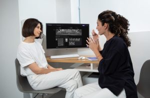
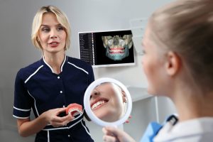
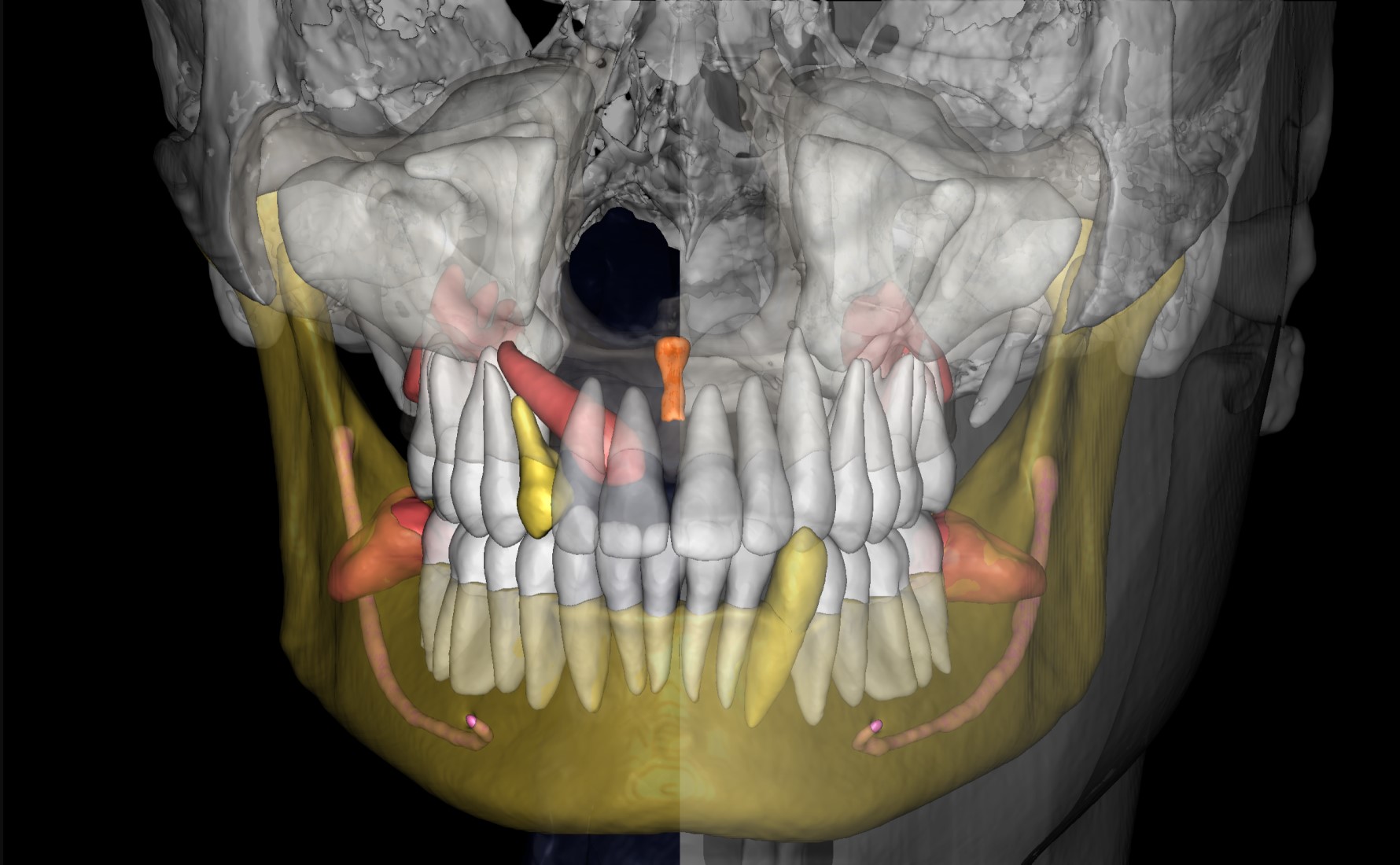
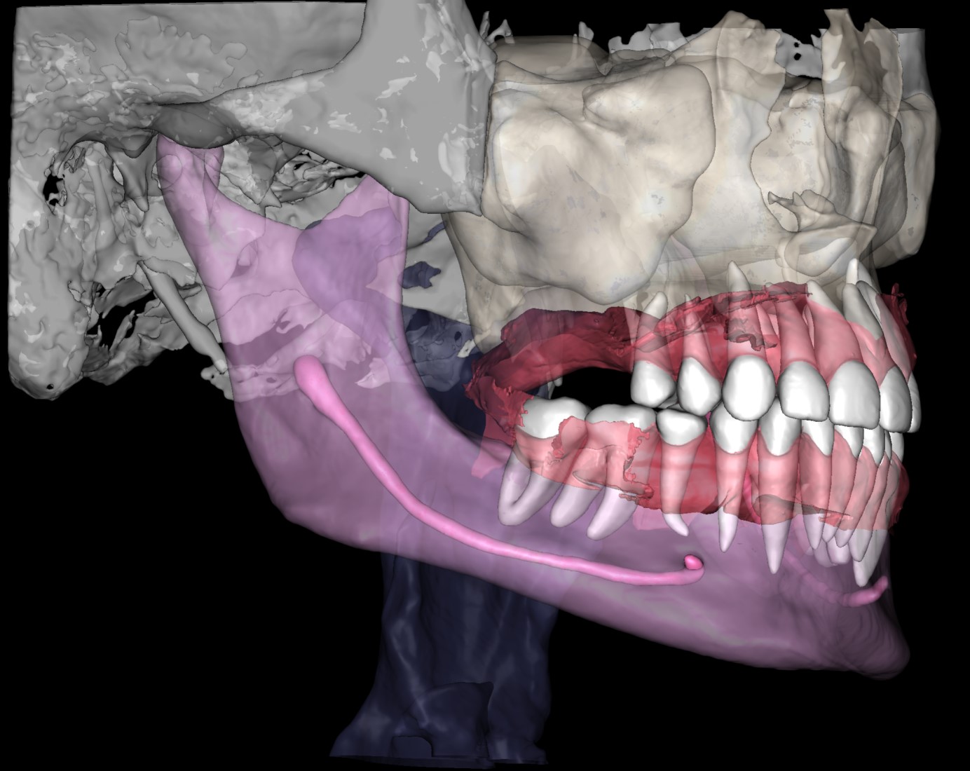
Includes fully automated segmentation and generation of 3D STL models, alongside simultaneous segmentation of various structures like soft tissue, teeth, maxillary sinus, inferior alveolar canal, incisive canal, mandible, maxilla, cranial base, and airways.
Generating 3D models with Diagnocat serves as an initial step for subsequent planning and modeling within specialized software or for 3D printing purposes.
Utilize the STL models for digital treatment planning, clinical case presentations, and engaging patients with effective communication, as visualizing the maxillofacial area significantly improves patient comprehension.
Who can benefit from this service?
Dental specialists
Diagnocat AI generates models of the upper and lower jaws, serving as digital counterparts to the models traditionally handled by doctors. The tool can also extract individual teeth from the model, enabling detailed planning for guided implant surgery.
Surgeons
The segmentation process encompasses all anatomical components, providing their volumes as STL files. Surgeons receive files for both upper and lower jaws, along with individual files for each tooth. This facilitates auto transplantation planning, augmentations, and the design of individual implants or membranes. It is worth noting that the cranial base bones remain undivided, presented as a unified structure within the model.
Orthodontist
Using the precision of AI, Diagnocat will segment teeth without including the surrounding bone, ideal for modeling splints and aligners. With CBCT volumes possessing a sufficient field-of-view (FOV), it enables the segmentation of pharyngeal and laryngeal airways, as well as facial soft tissues.
STL use cases
#Orthodontist Case 1
Objective
Implementing a comprehensive treatment plan and effective interaction between the clinician and the patient.
Challenge
When discussing the clinical situation, the orthodontist uses plaster models of the jaws, which are often complicated to understand for the patient.
Solution
As an alternative to traditional jaw models, Diagnocat suggests using 3D segmentation, generated based on cone-beam computed tomography (CBCT). In doing so, the patient can see their own face, teeth and bone. The viewing program increases the transparency of the soft tissue and shows the patient the pathology clearly. A visual 3D model simplifies communication, helps the patient understand the problem, and helps to set the patient up for timely treatment.
#Surgeon Case 1
Objective
To plan the implant placement with the use of a surgical guide.
Challenge
Manual anatomy segmentation, which requires a lot of time and professionalism of the clinician, especially if the quality of CBCT is low. Human factor and inaccurate superimposition of CBCT and IOS, which leads to errors in the creation of the surgical template.
Solution
Diagnocat automates CBCT segmentation, producing detailed 3D STL models. The process precisely segments full maxillofacial anatomy. With an intraoral scan, Diagnocat AI merges CBCT and IOS data for a unified model, widely used in digital dentistry treatment planning. The report is generated in just 5 minutes, saving your precious time.
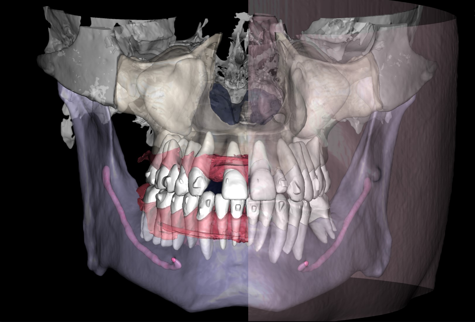
#Orthodontist Case 2
Objective
To reduce the amount of time taken to create a treatment plan for aligners.
Challenge
When planning tooth movements, the orthodontist needs a significant amount of time to evaluate the clinical case and perform calculations to achieve the most functional and accurate final tooth position.
Solution
Diagnocat's reports, based on CBCT and intraoral scans (STL files), help the clinician to quickly and accurately make decisions about treatment tactics and final tooth position, and to plan comprehensive treatment according to the individual needs of the patient.
#Surgeon Case 2
Objective
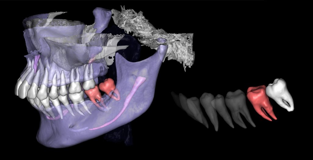
Establish communication with the patient and plan the autotransplantation of the third molar into the position of tooth 37 (Universal 18).
Challenge
The patient has doubts about the necessity of tooth extraction.
Solution
The automated process of segmentation and formation of 3D models from DICOM files allows extracting individual structures for subsequent 3D printing. The printed model of the third molar, taken from the "STL" module of Diagnocat, is used to prepare the socket for the transplanted tooth. The 3D reconstruction generated using Diagnocat displays the structure of the jaws and teeth and enables the visualization of tooth 37 (Universal 18) with periapical lesion around the roots. In this case, Diagnocat serves as a communication tool that helps convince the patient of the importance of timely implementation of the proposed treatment plan.
