Careful planning is crucial for reducing the chance of injury to the inferior alveolar nerve during lower third molar extraction
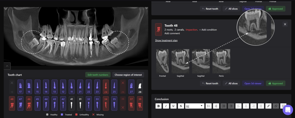
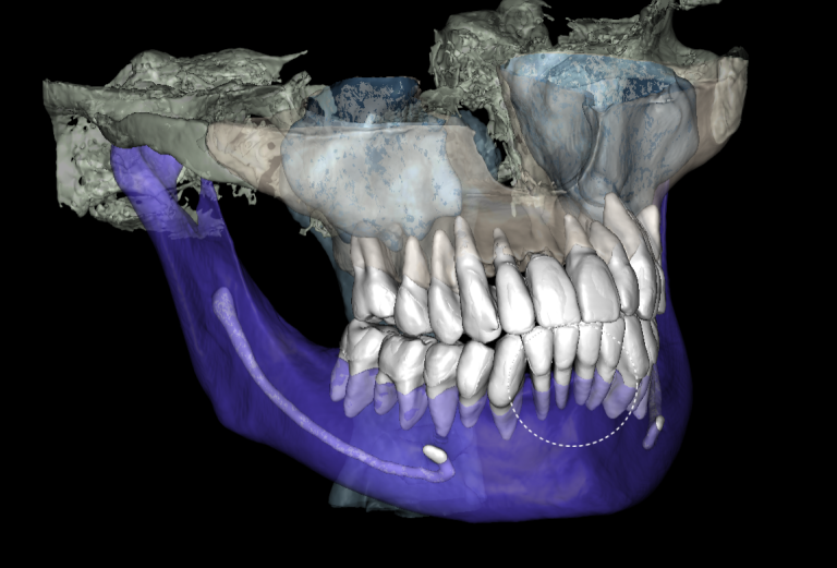
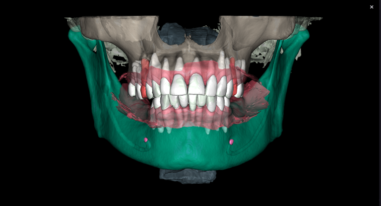
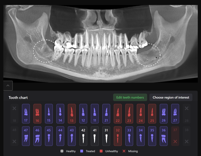
This clinical case is an example of how impacted mandibular third molars can be located in close proximity to the mandibular canal

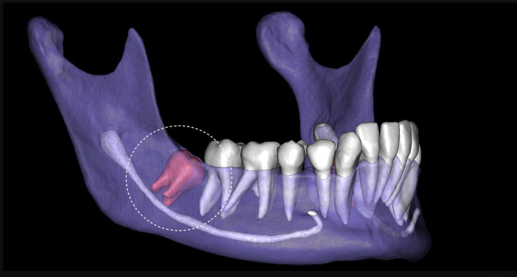
“Third Molar Report” created by Diagnocat AI is a tool, which provides accurate tracing of the mandibular canal, creates optimal 3D visualization and helps the clinician to determine the distance to the mandibular canal
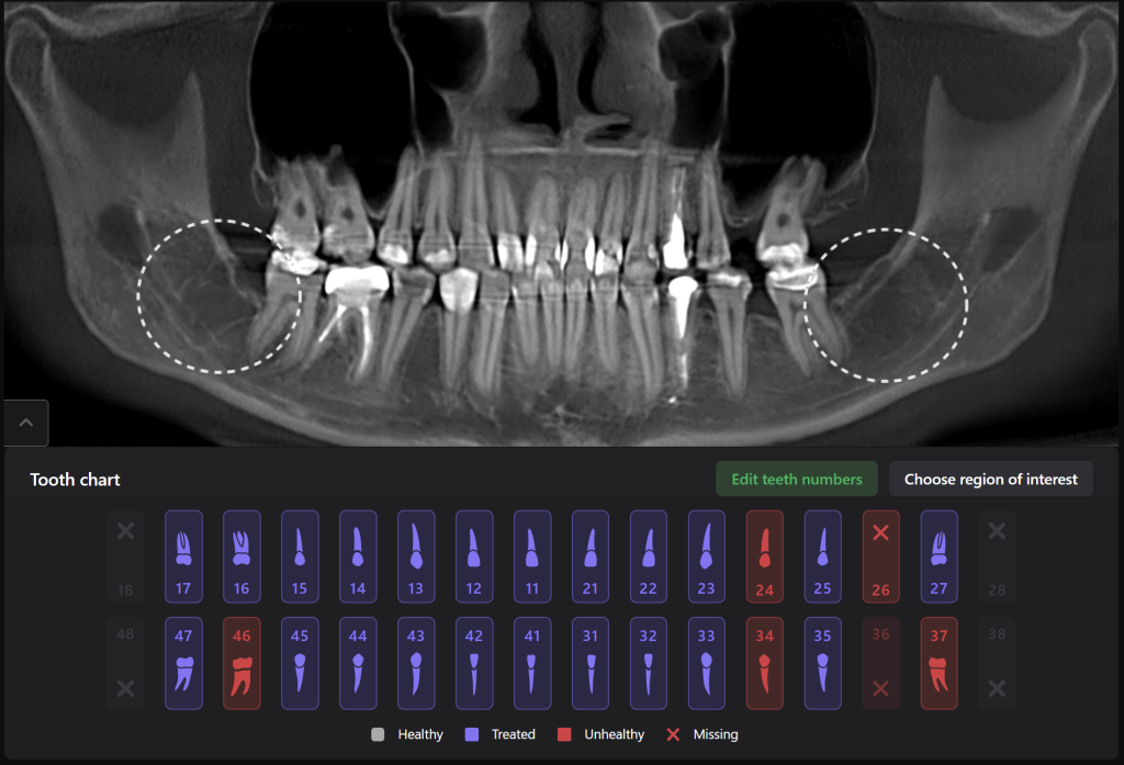
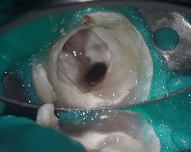
Teeth 38 (Universal 17) and 48 (Universal 32) were extracted with minimal surgical trauma, and without causing damage to the inferior alveolar nerves
