Dental Imaging Software—An Introduction
The operations of a dental practice have significantly changed over the years. In addition to innovative concepts and the latest dental materials, technology has become an indispensable ally in patient care. Among the most notable tech advancements, one that truly stands out is Dental Imaging Software.
The mouth is a small, inaccessible area of the body. Hence, a lot of the examination, diagnosis and treatment planning for oral diseases rely on radiographs that help dentists view the teeth, gums and oral tissues better. Dental Imaging Software is a digital platform that brings together all of the different threads of imaging in dentistry—acquiring, editing, reading, and sharing X-rays, CT scans, and images of intraoral cameras. Additionally, they allow treatment planning and provide easy-to-understand reports for patient communication.
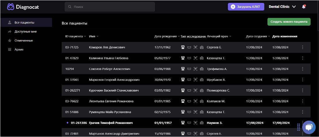
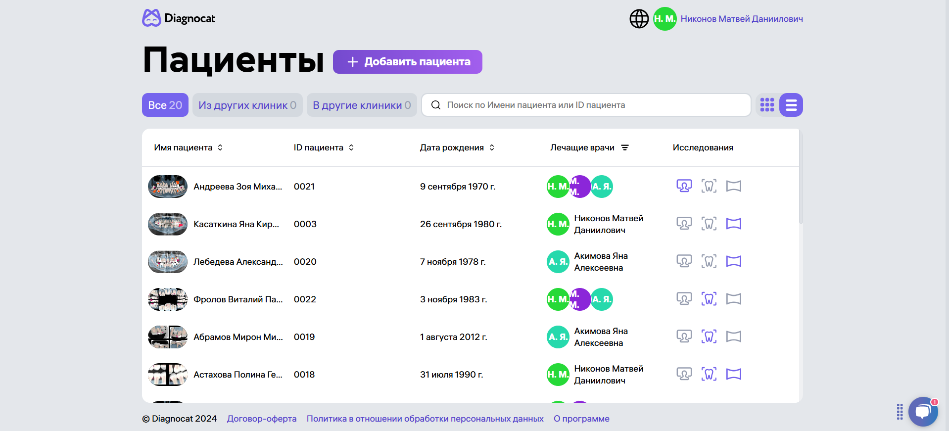
State-of-the-art dental imaging software today, like Diagnocat AI, uses artificial intelligence (AI) to analyse radiographs and scans [1]. This opens doors to new and more powerful dental imaging techniques that offer an incredibly accurate look at a patient’s oral health, helping dentists make more informed decisions [2].
Core Features of Dental Imaging Software
The key features of typical imaging software in dentistry are:
- High-resolution images: Dental imaging softwares is designed to capture high-definition, detailed images of hard and soft tissues of the mouth and reveal details not visible in traditional X-ray plates. These images help in detecting early-stage dental problems like occult caries (hidden cavities), vertical bone loss in the molars, a bony lesion, etc.
- 3D visualisation of radiographs and scans of the patient’s teeth, gums, and bone structure: This is critical for dental implant planning, orthodontic tooth alignment and complex yet minute procedures like root canal therapy (RCT).
3D visualisation is further equipped with two features: superimposition and segmentation.
Segmentation is the process of dividing an image into distinct segments or regions to isolate specific anatomical features (such as teeth, bones, soft tissues, and nerves) for focused analysis and diagnosis.
Superimposition allows dentists to compare two or more images that were captured at different times or under different conditions by overlapping them. This technique revolutionises the tracking of variations in a dental disease over time. Superimposition and segmentation often work in tandem to provide in-depth analyses.
- Image enhancement and measurements: These tools allow dentists to adjust contrast, brightness, and sharpness to detect hidden issues; they can also automatically measure important parameters such as tooth and bone height, width, and angulation for accurate treatment planning.
- Integration into practice management systems: They can be integrated into the practice management systems of the clinic, providing a holistic view of the patient’s dental health history alongside.
- Cloud storage and access: They also offer cloud-based storage, making it easier for dental teams to access images from any location—an ideal feature for practices with multiple locations.
- Sharing and multispecialty care: All records in dental image software can be shared as digital files and downloaded as raw image files, PDFs, or the desired formats. This is game-changing for dental referrals and multispeciality treatment planning.
Benefits of Imaging Software in Dental Practice
Adopting dental imaging software for your practice is not just about keeping up with trends in dentistry—it offers a host of tangible benefits, such as:
- Accuracy of diagnosis and streamlined treatment planning with less scope for errors and operational surprises.
- Faster diagnosis; software such as Diagnocat AI has the ability to read over 65 dental conditions on CBCT images, including rare pathologies and non-dental findings like sinus issues and bone structure deformations, as well as 35 conditions in 2D images, bitewings, FMX, and panoramic radiographs.
- Easier inter-specialty collaboration
- Easy patient communication; better patient retention and conversions.
- Reduced overheads and cost-efficiency
When combined, these advantages improve patient satisfaction and clinical results [3].
How to Choose a Dental Imaging Software
Dental imaging software is not just about taking radiographs; it’s about transforming the way these images are processed, analysed, and utilised for effective treatment planning. The points below will help decide which dental imaging software to invest in:
- The core features of the product must match the needs of the dental practice: For example, implant dentists and orthodontists need extensive virtual treatment planning tools, preview features, and user-friendly reports for patient communication.
- Compatibility with the existing imaging equipment in the practice and patient management systems is also crucial for seamless operation.
- Scalability: It is best to choose a system that can scale with your practice. Cloud-based solutions are particularly beneficial for practices with multiple locations or those planning to expand.
- Cost, regulatory compliance, and other specifics: The software must adhere to the regional health regulations, fit within the practice’s budget and have flexible subscription plans for ease of use.
- Customer support: A reliable customer support team is a must; the software provider must also offer regular updates to enhance performance going forward.
Leading Software Options in Dentistry
Amongst the others, Diagnocat AI is a prominent player in the dental imaging software industry. It is also the first dental imaging software to provide AI analysis features. Several other dental imaging software options stand out in the market owing to their performance, dependability, and user-friendly features; you can select any of them based on your practice’s location, budget, and requirements.
Future Trends in Dental Imaging
As technology continues to evolve, dental imaging software has incorporated more advanced features over the past decade. As previously mentioned, Diagnocat AI was one of the pioneers in integrating AI into dental imaging techniques—a transformative way to detect early signs of cavities, periodontal issues, and oral cancers—and assists in predicting treatment outcomes, enabling dentists to make more informed decisions.
Other future possibilities in digital dental imaging could be the integration of Augmented Reality (AR) in imaging and adding the 4D aspect to imaging—the element of time—to visualise dynamic processes like jaw movements and temporomandibular joint (TMJ) disorders, etc.
FAQ
- How is dental imaging software applied in the dental setting?
Dental imaging software can be used in a dental setting to acquire radiographs, and 3D scans, analyse, prepare reports, store records, share files and help in treatment planning.
- How much does dental imaging software cost?
The cost of dental imaging software varies depending on the features, the type of imaging technology (e.g., 2D, 3D), and the vendor. It’s important to consider both upfront and ongoing costs, including training, support, and maintenance.
- How does dental imaging software reduce radiation exposure?
Dental imaging software, especially digital radiography systems, uses less radiation compared to traditional X-ray methods. The images are captured quickly and processed digitally, requiring less exposure to achieve high-quality results.
References:
- https://www.oralhealthgroup.com/features/diagnocat-the-first-and-only-imaging-ai-in-3d-comes-to-dentistry-and-canada/
- De Angelis F, Pranno N, Franchina A, Di Carlo S, Brauner E, Ferri A, Pellegrino G, Grecchi E, Goker F, Stefanelli LV. Artificial Intelligence: A New Diagnostic Software in Dentistry: A Preliminary Performance Diagnostic Study. Int J Environ Res Public Health. 2022 Feb 2;19(3):1728. doi: 10.3390/ijerph19031728. PMID: 35162751; PMCID: PMC8835112.
- Shujaat S, Bornstein MM, Price JB, Jacobs R. Integration of imaging modalities in digital dental workflows – possibilities, limitations, and potential future developments. Dentomaxillofac Radiol. 2021 Oct 1;50(7):20210268. doi: 10.1259/dmfr.20210268. Epub 2021 Sep 14. PMID: 34520239; PMCID: PMC8474138.


