Specialists Reports*
*The availability of Diagnocat products is limited in various countries. Please contact us to check availability in your country.
Diagnocat offers a range of specialist reports including the third molar, orthodontic, implantology, and endodontic reports, easily accessible through the platform.
Third Molar Report
Third molar (“wisdom tooth”) extraction is often necessary for reasons related to impaction, ectopic position, or planned orthodontic and restorative treatments. However, this surgery involves risks due to the tooth’s unusual shape and proximity to critical anatomical structures like the inferior alveolar nerve and maxillary sinus. This necessitates precise diagnosis and visualization during the planning phase.
Diagnocat’s AI accurately traces the inferior alveolar nerve canal, generating a report with clear cross-sectional slices illustrating its proximity to the wisdom tooth. This report assists the clinician in understanding extraction risks, selecting the appropriate surgical approach, and effectively explaining the situation to the patient.
Report generation within 3 minutes.
Features an extensive range of cross-sectional slices for precise clinical assessments.
Produces accurate tracing of the mandibular canal.
Facilitates the surgical process by identifying risks, improving planning, and enabling effective patient communication.
Detailed and patient-friendly report available for printing or download in PDF format.
Orthodontic Report
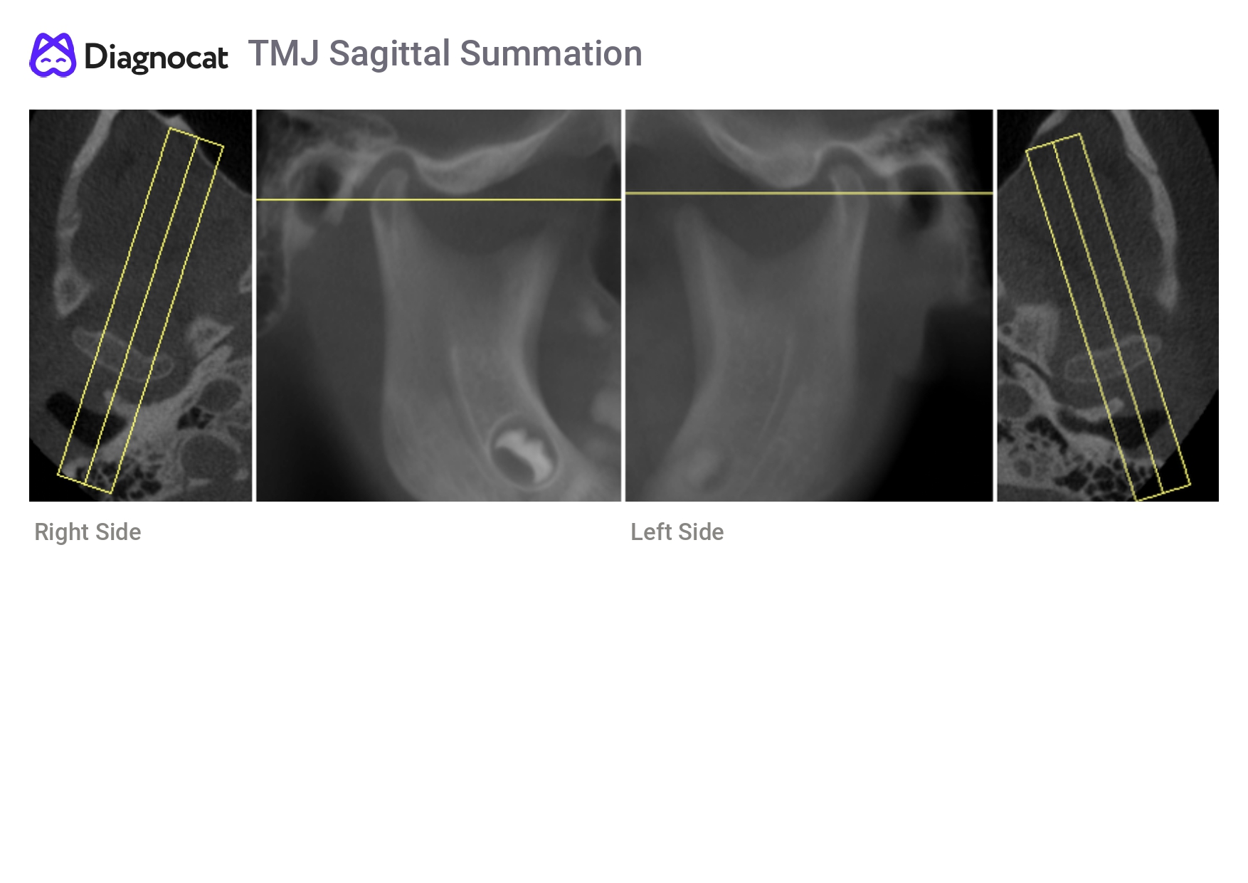
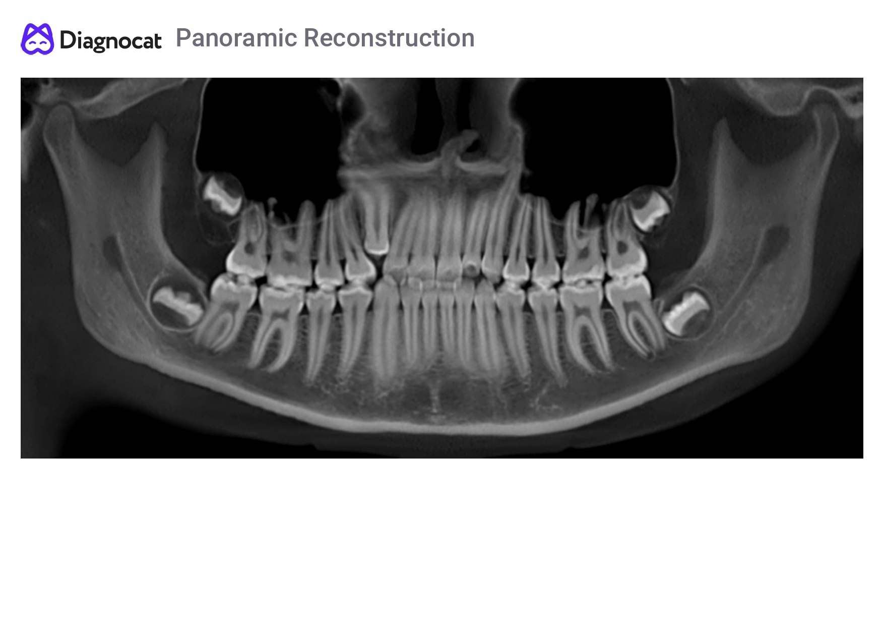
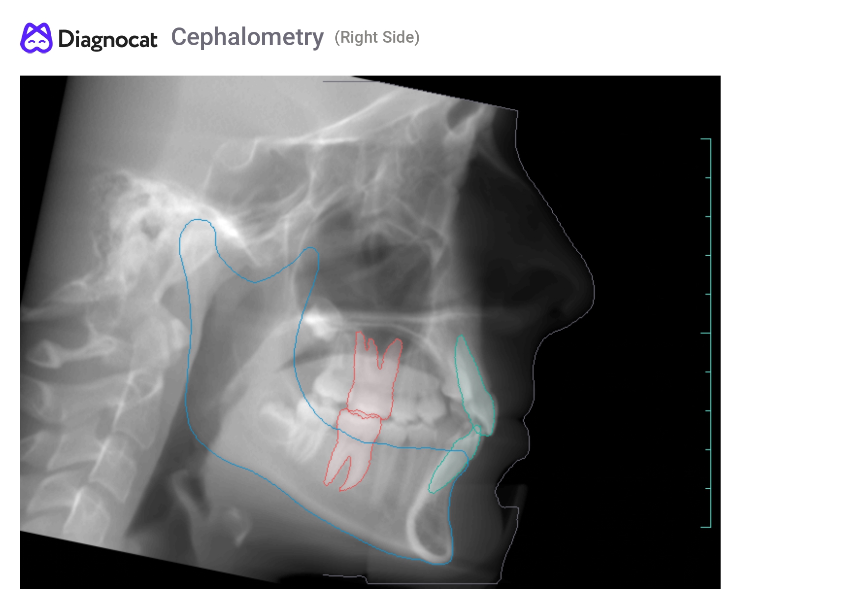
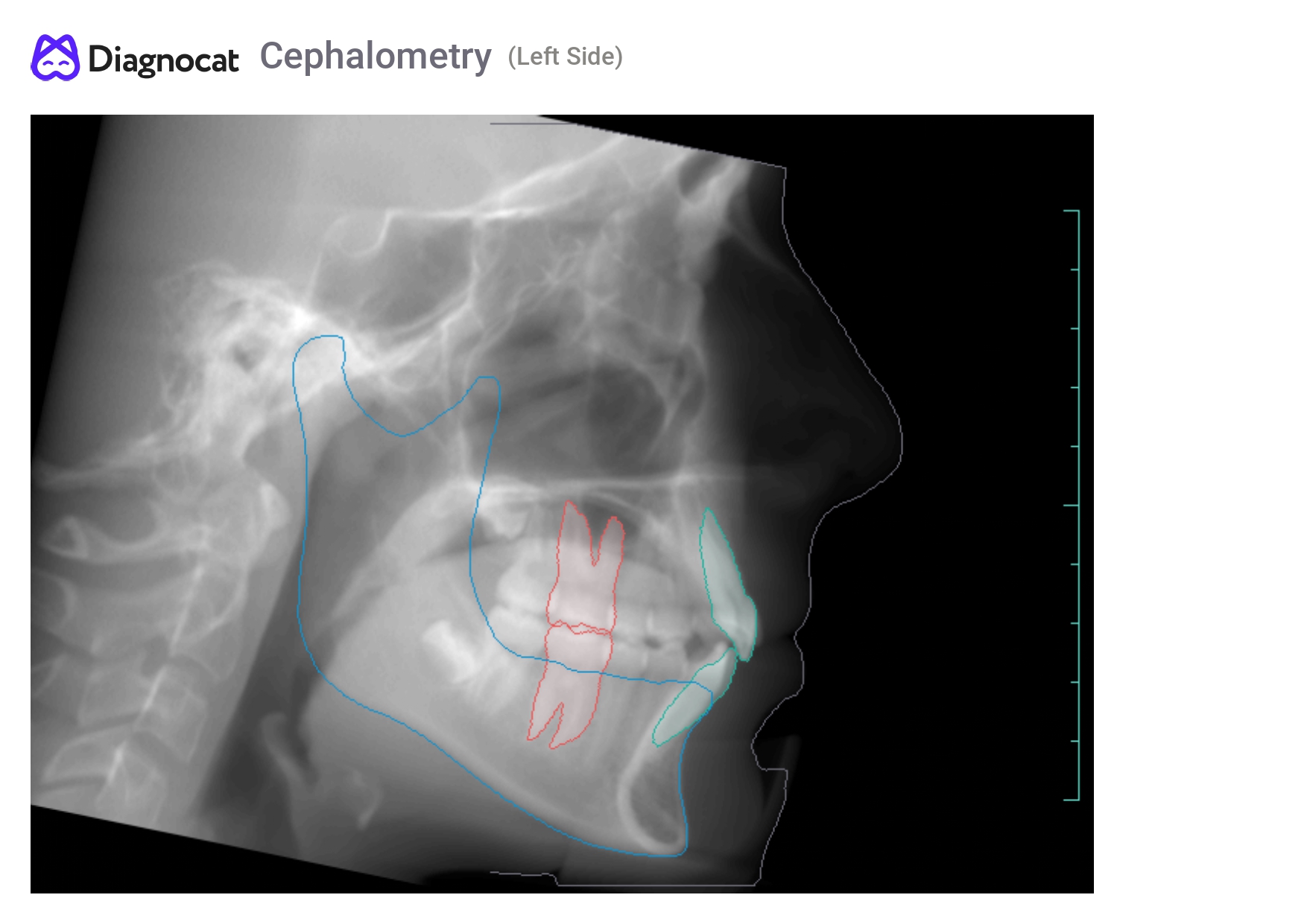
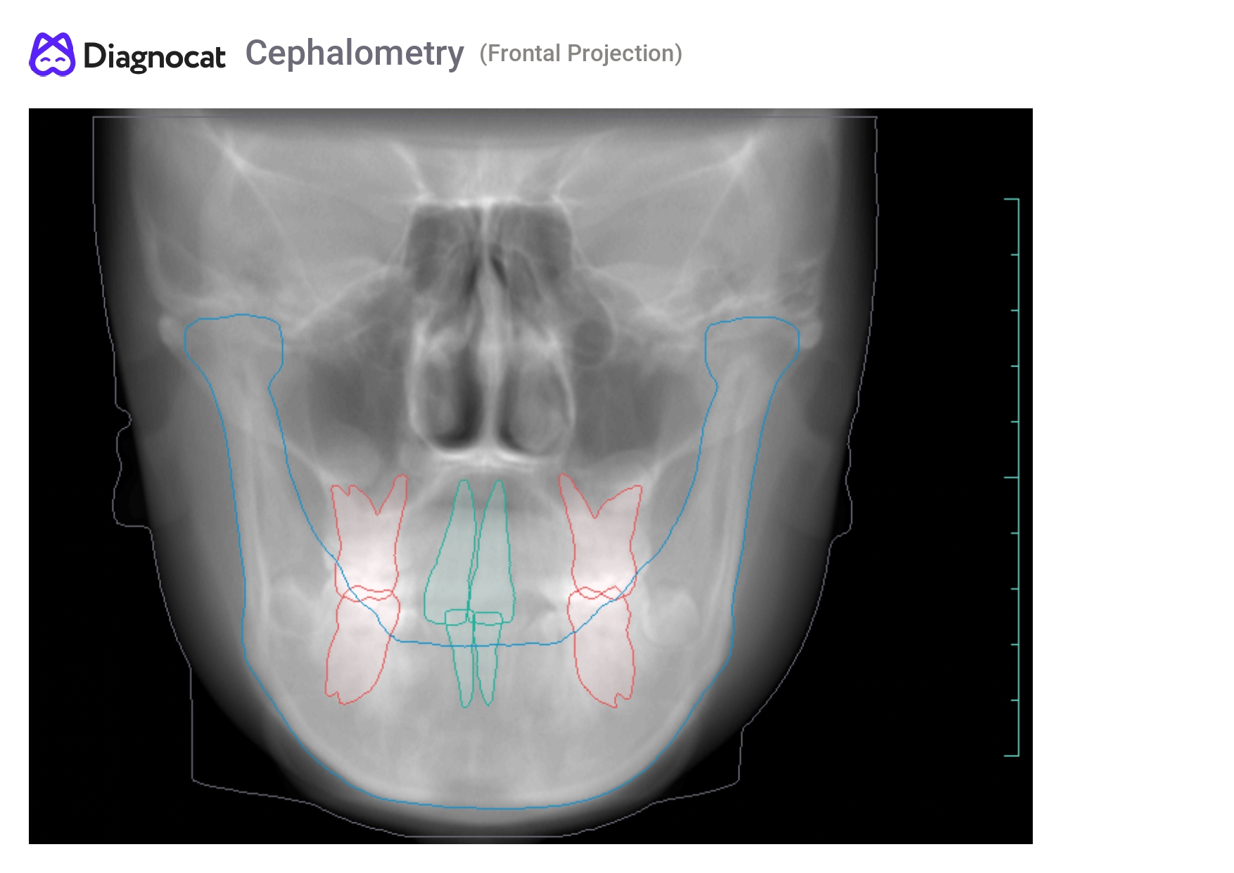
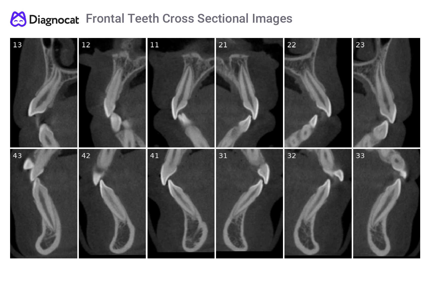
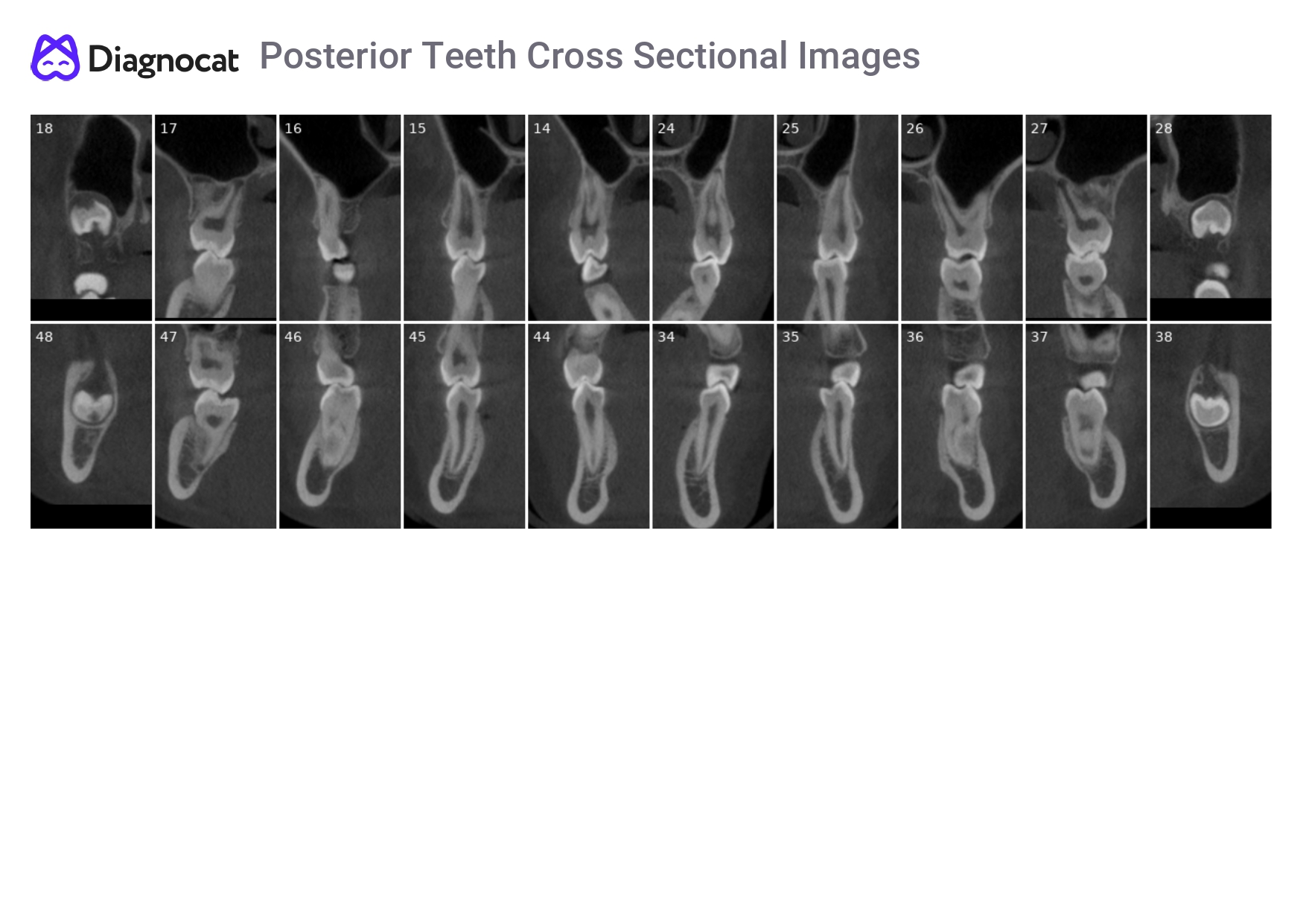
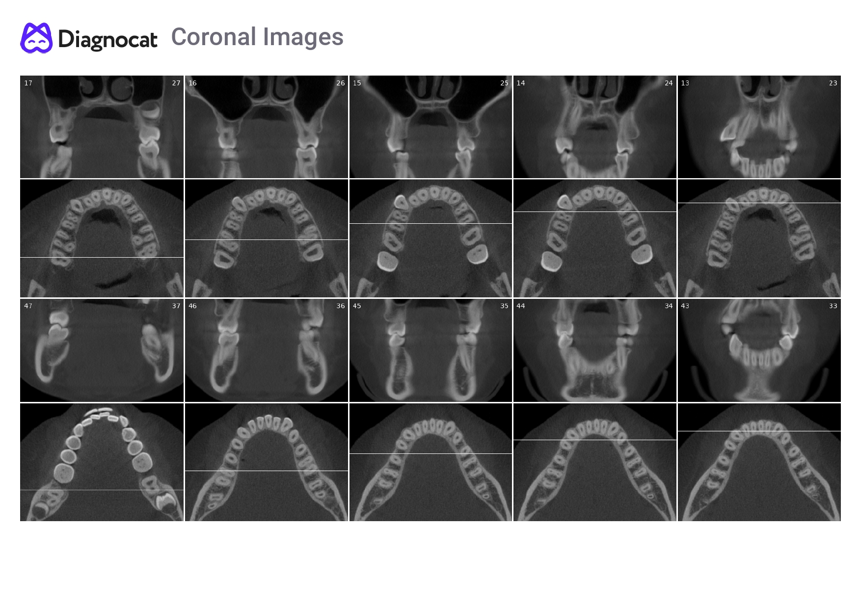
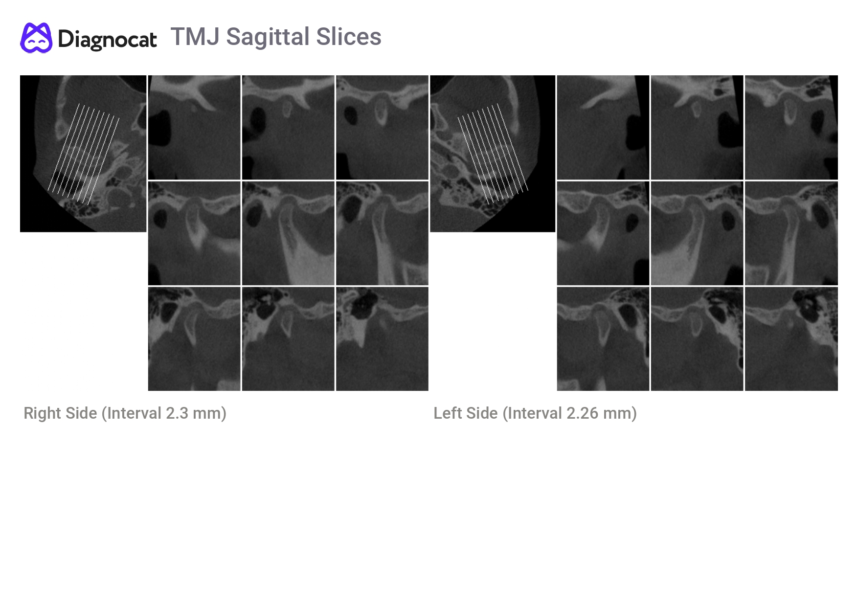
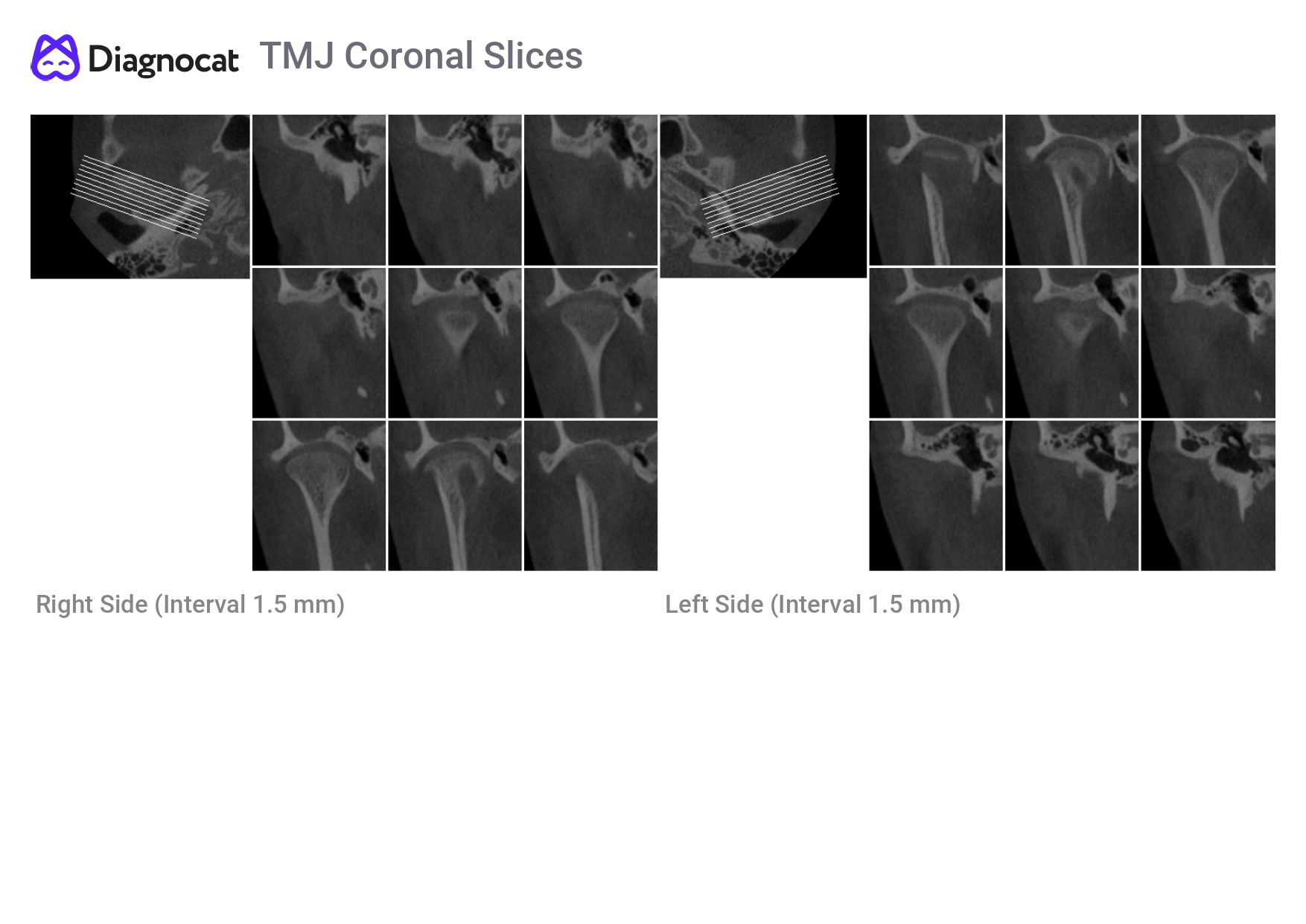
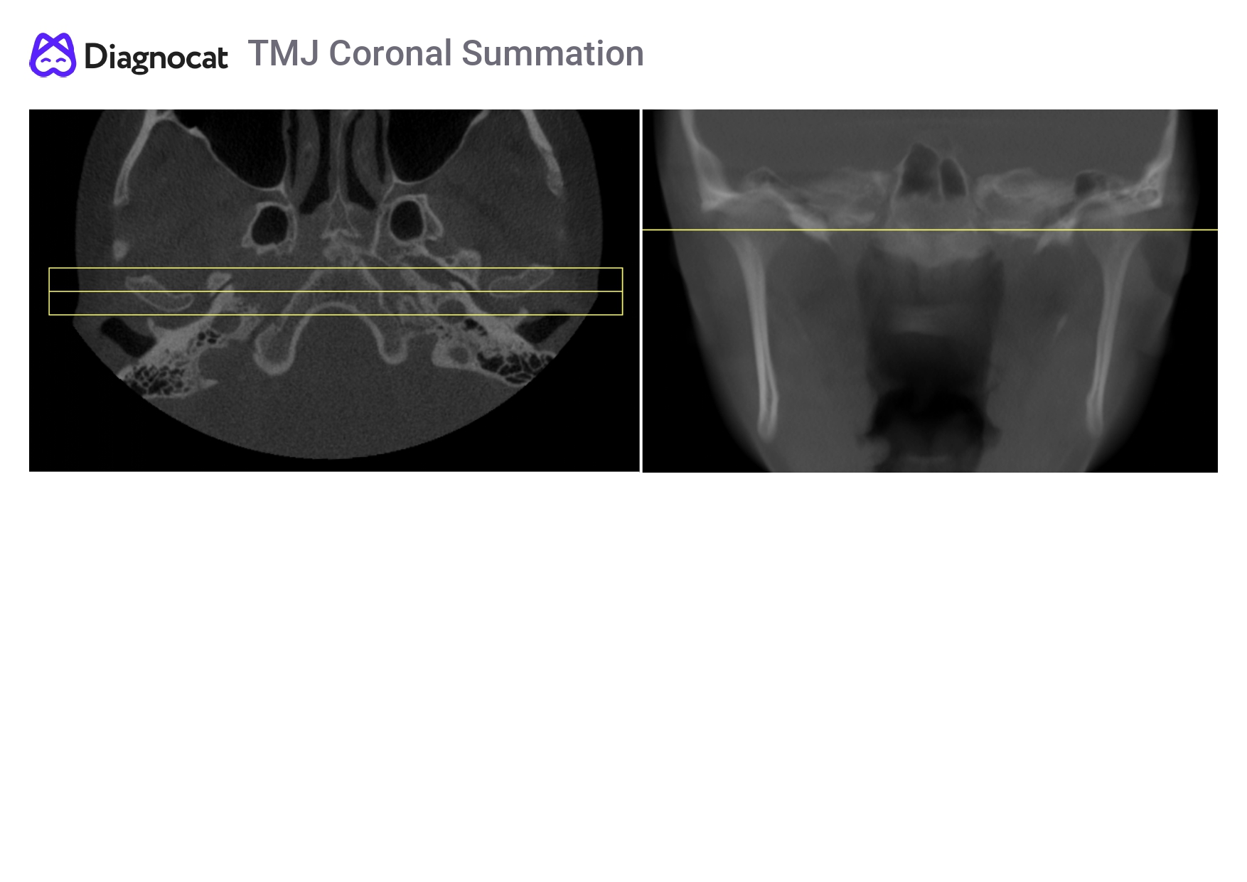


Diagnocat’s orthodontic report provides a comprehensive analysis of the patient`s dentofacial system using CBCT data.
Advantages of the Diagnocat orthodontic report:
Report generation within 5 minutes.
Clear visualization of the TMJ for easy assessment.
Detailed and patient-friendly report available in printable or downloadable PDF format.
The Diagnocat orthodontic report includes:
Panoramic CBCT reconstruction
Cephalometric reconstructions (right, left, and frontal)
Cross-sectional views of all teeth
Coronal slices of dental arches in molar and premolar areas
TMJ cross-sections and reformatted views
Implantology Report
Powered by the precision of AI, Diagnocat delivers a next generation experience in implantation planning reports for medical professionals. The implantological report includes a pan-like image reformatted from CBCT, providing an in-depth understanding of the patient’s clinical case. An extended set of cross-sectional slices provides detailed bone structure and configuration measurements, improving the predictability and success of the implantation process. Diagnocat meticulously analyzes relationships with critical anatomical structures like the maxillary sinus and mandibular canal, mitigating potential complications and ensuring procedural safety. The AI-generated implant report serves as a vital tool for dental professionals, guaranteeing quality and personalized patient care. This advancement raises the bar in implantology practice, resulting in increased success rates while meeting the needs and expectations of professionals and patients alike.
This service features:
Report generation within 3 minutes
Panoramic reformatting from CBCT for comprehensive patient case understanding
Detailed cross-sectional slices with bone structure
Customizable slice thickness
Detailed and patient-friendly report available in printable or downloadable PDF format
Endodontic Report
Our Endodontic Report serves as a valuable tool for diagnosing and planning root canal treatments. Powered by AI, Diagnocat generates a comprehensive report detailing the root canal system’s shape and diagnoses the furcation area. Accurate measurement of periapical pathology aids the dentist in seamlessly evaluating the treatment process. Moreover, the AI highlights periapical lesions, providing patients with a clear and understandable visualization.
This service features:
Report generation within 3 minutes
Detailed AI-generated cross-sectional slices depicting the root canal system and furcation area
Accurate measurement of periapical pathology for unbiased endodontic treatment evaluation
AI-generated visuals aiding patient comprehension of periapical lesions
Customizable slice thickness and intervals
Detailed and patient-friendly report available in printable or downloadable PDF format
Explore more diagnocat products
Diagnocat presents a range of solutions tailored to the different needs of your dental practice.
Cloud storage and Viewer
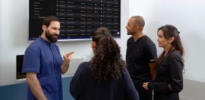
All dental images and reports are securely stored in your cloud-based personal account,
accessible for viewing, uploading, sharing, or printing from any device.
Collaboration Tool*
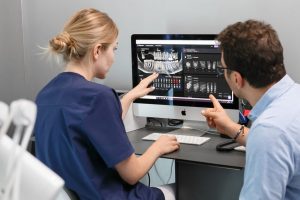
Introducing Diagnocat’s Platform for Comprehensive Treatment Plan Management that
transforms our AI into your virtual dental clinic assistant.
Superimposition
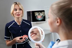
Our superimposition feature offers dental specialists an enhanced view of their patient's oral
cavities by combining the advantages of CBCT and intra-oral imagery.
Radiology Report
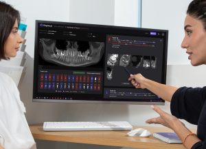
Diagnocat’s AI analysis of intraoral X-rays, panoramic X-rays (OPGs), and CBCT images produces an accurate, clear, and concise report of over 65 conditions.
CBCT Segmentation
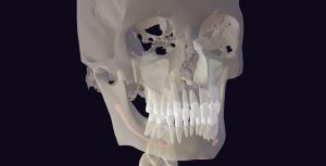
Diagnocat AI's automatic segmentation feature transforms CBCT files into a 3D STL model, a
pivotal innovation for digital dentistry.
Cloud storage and Viewer

All dental images and reports are securely stored in your cloud-based personal account,
accessible for viewing, uploading, sharing, or printing from any device.
Collaboration Tool*

Introducing Diagnocat’s Platform for Comprehensive Treatment Plan Management that
transforms our AI into your virtual dental clinic assistant.
Superimposition

Our superimposition feature offers dental specialists an enhanced view of their patient's oral
cavities by combining the advantages of CBCT and intra-oral imagery.
Radiology Report

Diagnocat’s AI analysis of intraoral X-rays, panoramic X-rays (OPGs), and CBCT images produces an accurate, clear, and concise report of over 65 conditions.
CBCT Segmentation

Diagnocat AI's automatic segmentation feature transforms CBCT files into a 3D STL model, a
pivotal innovation for digital dentistry.
Cloud storage and Viewer

All dental images and reports are securely stored in your cloud-based personal account,
accessible for viewing, uploading, sharing, or printing from any device.
Collaboration Tool*

Introducing Diagnocat’s Platform for Comprehensive Treatment Plan Management that
transforms our AI into your virtual dental clinic assistant.
Superimposition

Our superimposition feature offers dental specialists an enhanced view of their patient's oral
cavities by combining the advantages of CBCT and intra-oral imagery.
