Radiology Report

Increasing treatment acceptance
Diagnocat AI produces a report that helps explain the detected conditions supporting a treatment plan in a simple, comprehensive manner. This report can be printed in the office or emailed, as a PDF to the patient. A clear, graphic summary of the results provides transparency and increases the treatment acceptence.
Quick and accurate
On average, it takes at least 30 minutes for a specialist in oral radiology to analyze a CBCT. Radiological findings of conditions are generated by Diagnocat within 10 seconds for 2D images and 4-6 minutes for CBCT images. These AI supported reports provide the professional with a quick screening tool for common dental findings.
Radiologic report nuances
What can be seen
What cannot be detected
Detection Statistics
CBCT images
Diagnocat automatically produces a patented, panoramic reformation of the CBCT. This panoramic reconstruction is a unique AI-driven feature that provides professionals with rapid and convenient case reviews.
The intuitive interface assists the clinician in navigating through the entire scan, highlighting previously treated teeth and newly identified pathologies. The clinician can evaluate the images in two ways:
Online 3D viewer - provides a multiplanar reconstruction of the specific tooth, allowing the clinician to rotate and align the viewer axes as necessary.
A complete series of automatically-generated cross sections.
CBCT Radiology Report advantages
Automatically-generated cross sections
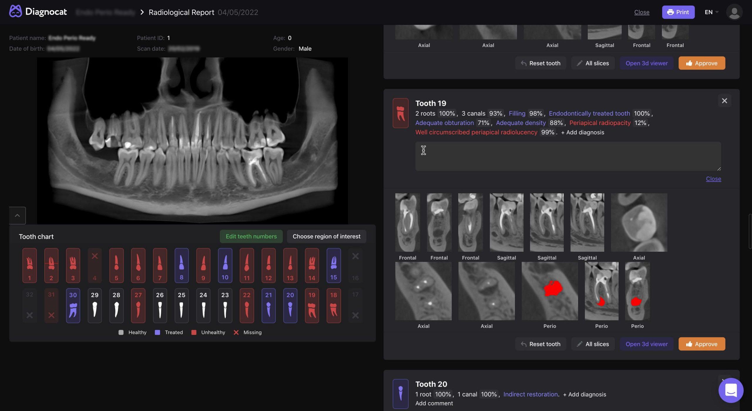
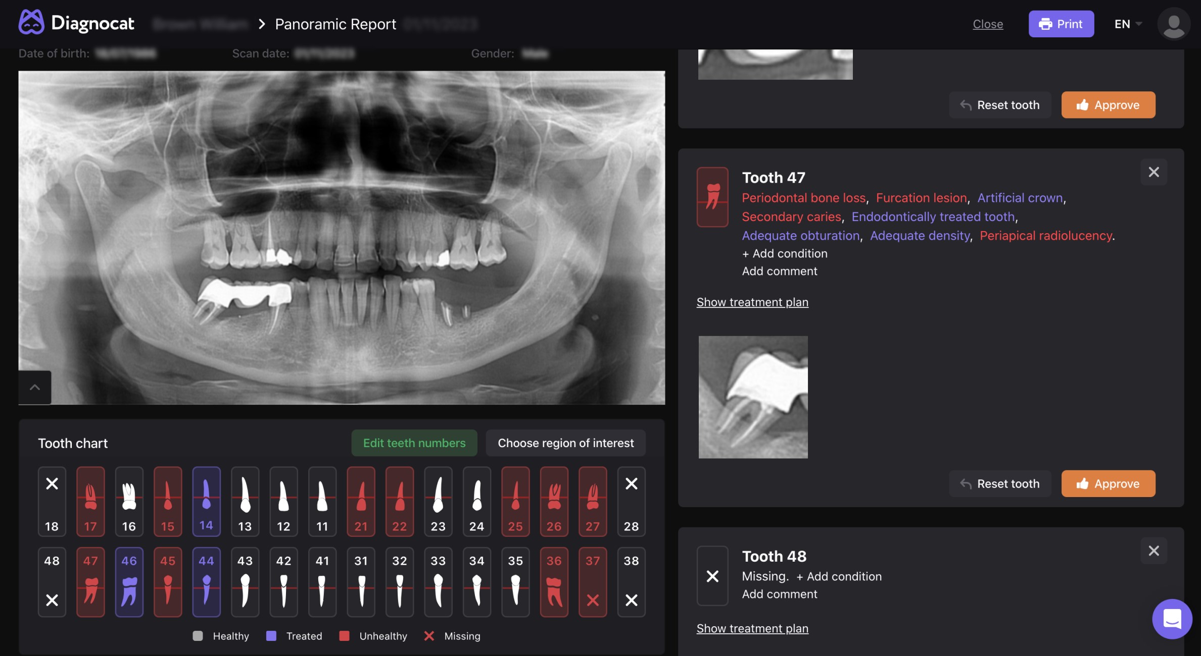
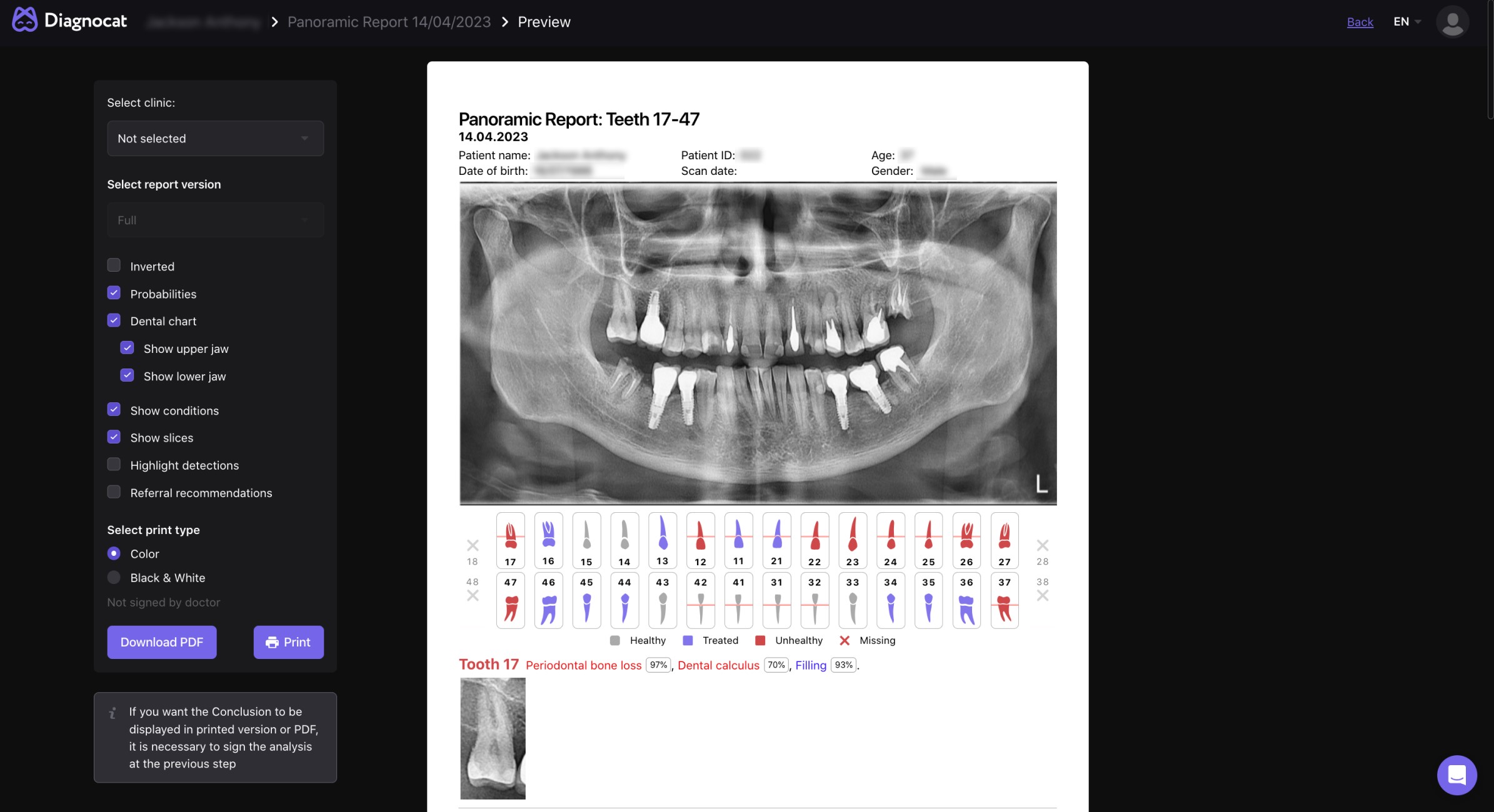
The dental chart navigates professionals to the region of interest, where the dentist interface will clearly display the detected conditions and the AI’s confidence in its recommendation (displayed in %).
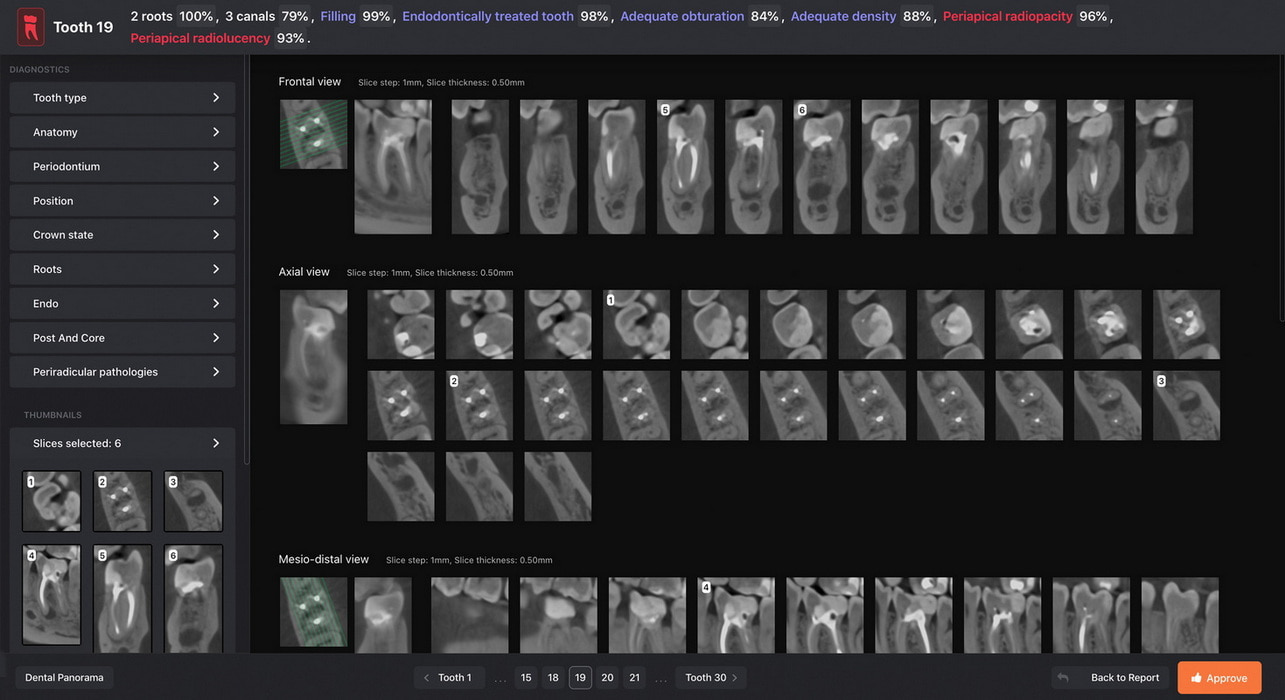
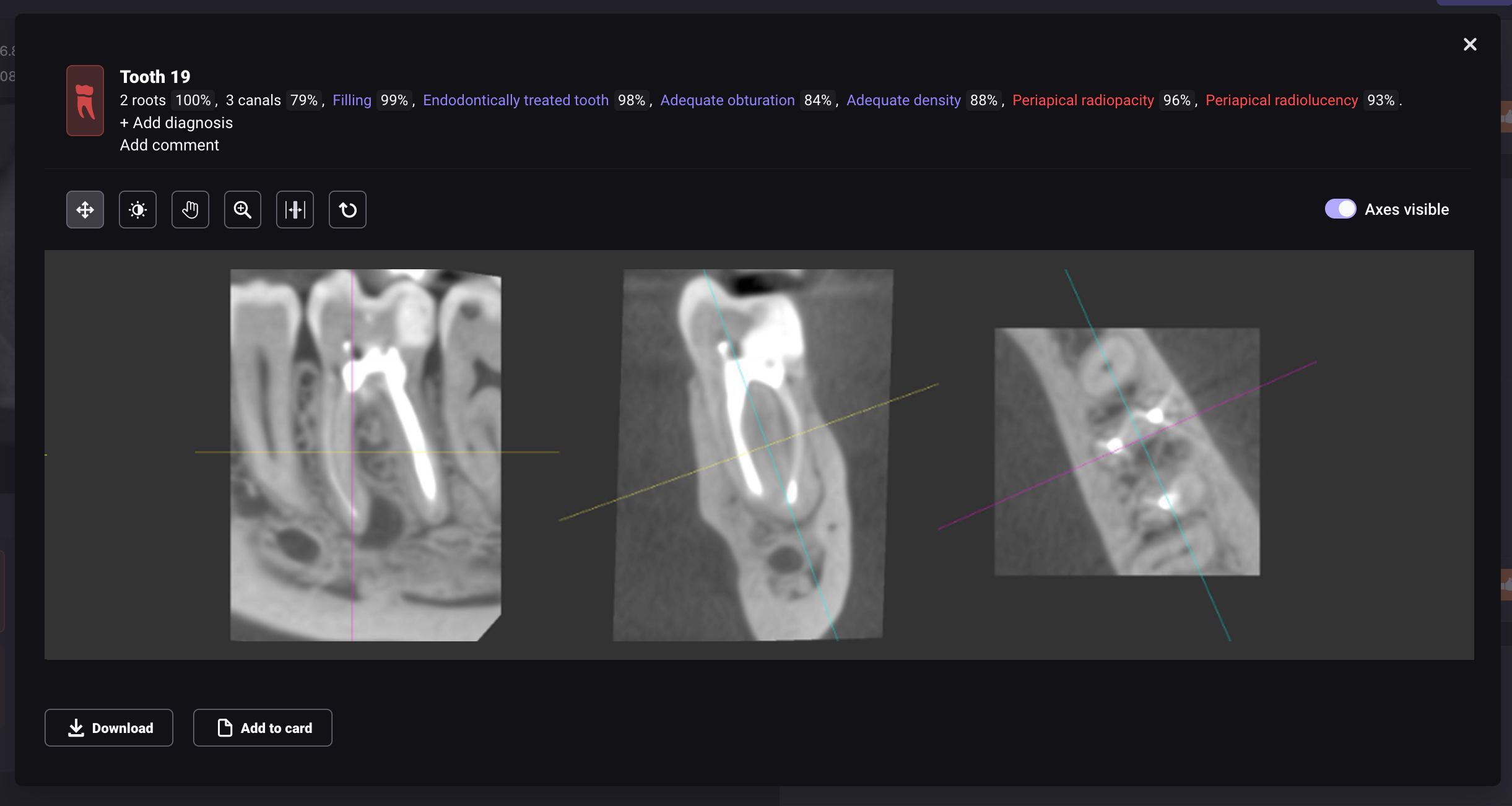
Online 3D viewer
To obtain more details about a tooth without returning to your imaging software, we’ve included two valuable functions: Multiplanar view (MPR), which confines the field of view to a single tooth volume. Diagnocat also features interactive axis movement for an in-depth analysis and the creation of tailored sectional views that can supplement the auto-generated sections. Reviewing previously generated tooth sections. Diagnocat AI produces a series of sectional images in primary projections. The algorithm may highlight potential problematic areas to help direct the dentist's attention.
Detailed Radiological Screening
After the clinician looks through Diagnocat’s suggestions, they can accept or reject them, add comments, and supplement the report with additional photos or images.
Printed Report
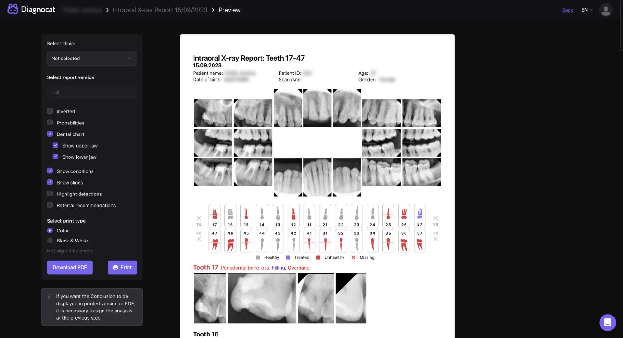
The next step is to generate a comprehensive report detailing findings and conditions. This report encompasses a dental chart with individualized descriptions for each tooth, conveniently packaged in a printable PDF file. Any teeth with detected conditions are highlighted in red, facilitating patient discussions. This report complements the treatment plan by establishing a direct link between the patient's conditions and the necessary treatments. Additionally, for easier comprehension, a simplified one-page report can be created, which features a reformatted panoramic view accompanied by a color-coded dental chart.
CBCT pre-filled report
A pre-filled report on dental findings for each tooth is confirmed with a few clicks. It includes pathologies and conditions indicated with specific colors on the dental chart.
Panoramic view
Automatic creation of a high-quality reformatted panoramic view from a CBCT volume
Dental chart
Color-coded dental chart – Showing healthy, previously treated, missing, and unhealthy teeth with treatable conditions
65+ conditions
The list of 65+ conditions for CBCT images, including sinus and bone structure abnormalities screening, periapical lesion, periodontal bone loss, signs of caries, impactions, and defects of previous endodontic treatment among many others to help dental professionals become more productive
Cross-sections
Automatic creation of typical cross-sections for each tooth in 3 separate projections
Multiplanar viewers
Focused multiplanar viewers for each tooth
Recommendations
Referral recommendations
PDF report
Production of a printed PDF report for the medical record and improved communication with your patient
Visualization
Graphical and volumetric visualization of periapical lesion
Detection
Initial detection of bone structures and maxillary sinus abnormalities
Intraoral X-ray
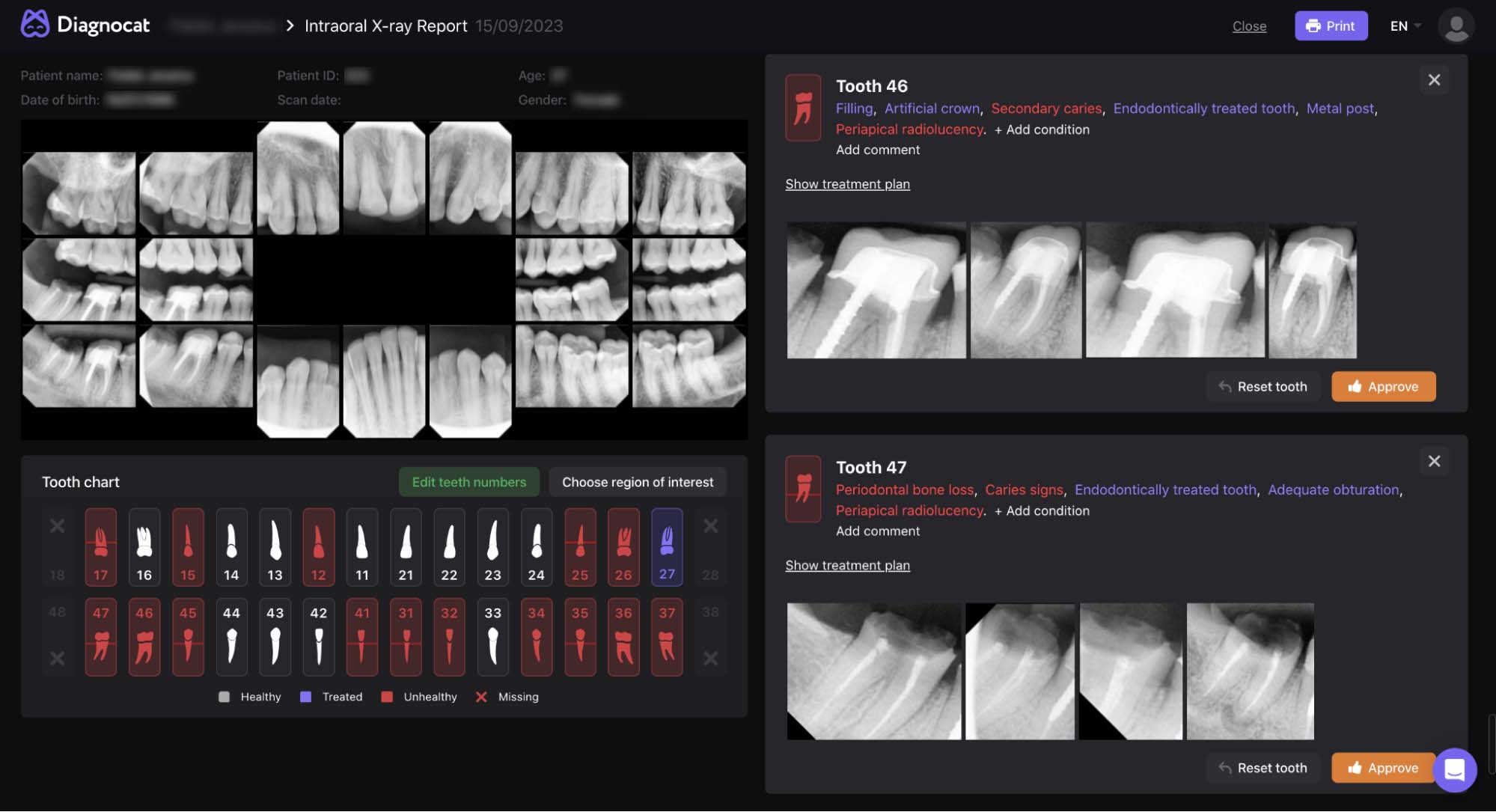
The AI analysis of intraoral X-rays, ranging from a single image to a full-mouth series of up to 24 images, automatically organizes the images into a simplified template before assigning numbers to the teeth and creating a radiological report detailing findings for each individual tooth.
Intraoral X-ray advantages
Detailed radiology report
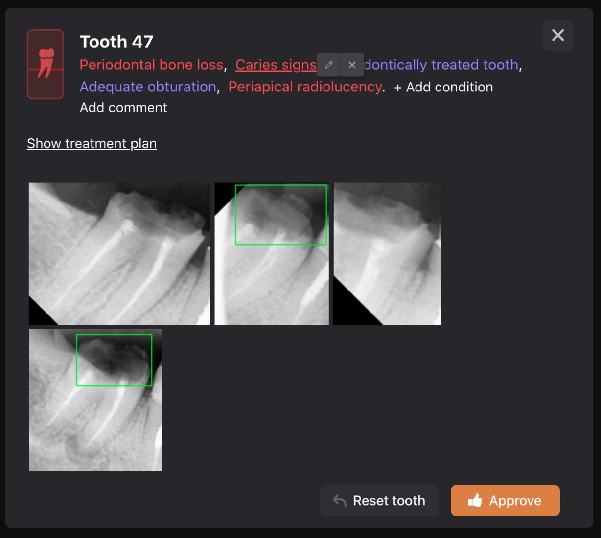
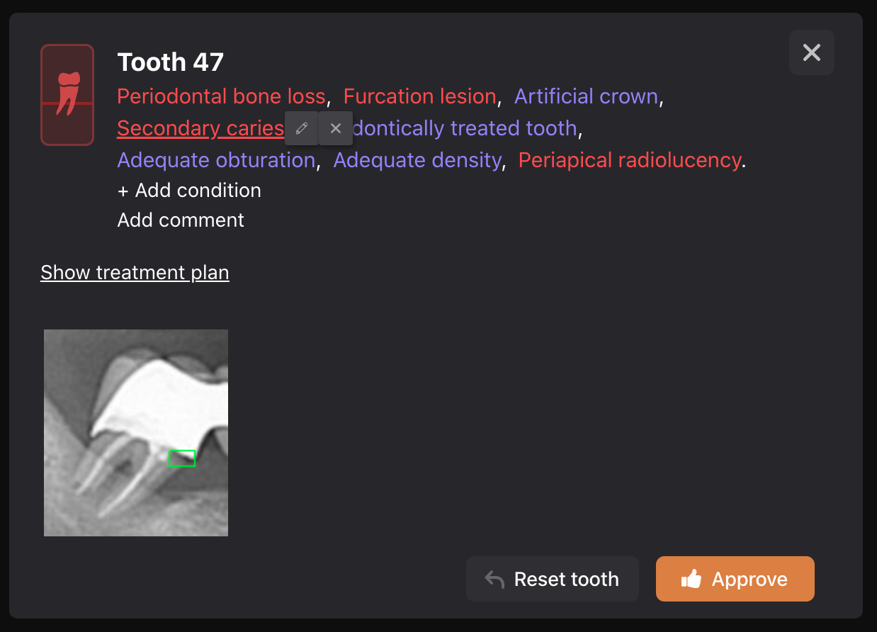
For a more effective examination of tooth conditions, Diagnocat offers a focused visualization of each tooth, allowing the dentist to concentrate on the region of interest.
Visualization and clinical case presentation
With the use of AI, radiological findings can be highlighted, making this tool useful for both diagnosis and patient communication.
Visual representation ensures clarity in the treatment process and increases patient trust.
Printed report
The report is generated in PDF format and can be emailed or printed.
Teeth in need of treatment are highlighted in red, which is especially convenient for discussing the report with the patient.
A detailed radiological report complements the treatment plan and allows the patient to visually understand the connection between oral health and necessary treatment.
Intraoral X-ray pre-filled report
Image visualization
Image visualization with convenient processing tools
Dental findings
AI-generated dental findings
Dual frames
Dual frames highlighting detected conditions on an image
Partial view
Partial view of intraoral X-rays focused on specific teeth
PDF report
Production of a printed PDF report for the medical record and improved communication with your patient
25+ conditions
The list of possible findings consists of 25+ different conditions and includes signs of caries, dental calculus, periapical lesion, periodontal bone loss, visible defects of previous endodontic treatment, lack of interproximal tooth contact and others designed to improve the efficiency of dentists
Panoramic radiograph (OPG)
The Diagnocat algorithm produces a report with AI-generated findings based on the panoramic image your office uploads.
OPG advantages
Detailed radiology report
For a more efficient tooth conditions analysis, Diagnocat offers visualization for each individual tooth, allowing doctors to concentrate on specific regions of interest.
Visualization and clinical case presentation
Artificial intelligence highlights important radiological findings, making this tool useful for both diagnosis and patient communication. Visualization enhances transparency in the treatment process and helps build trust with the patient.
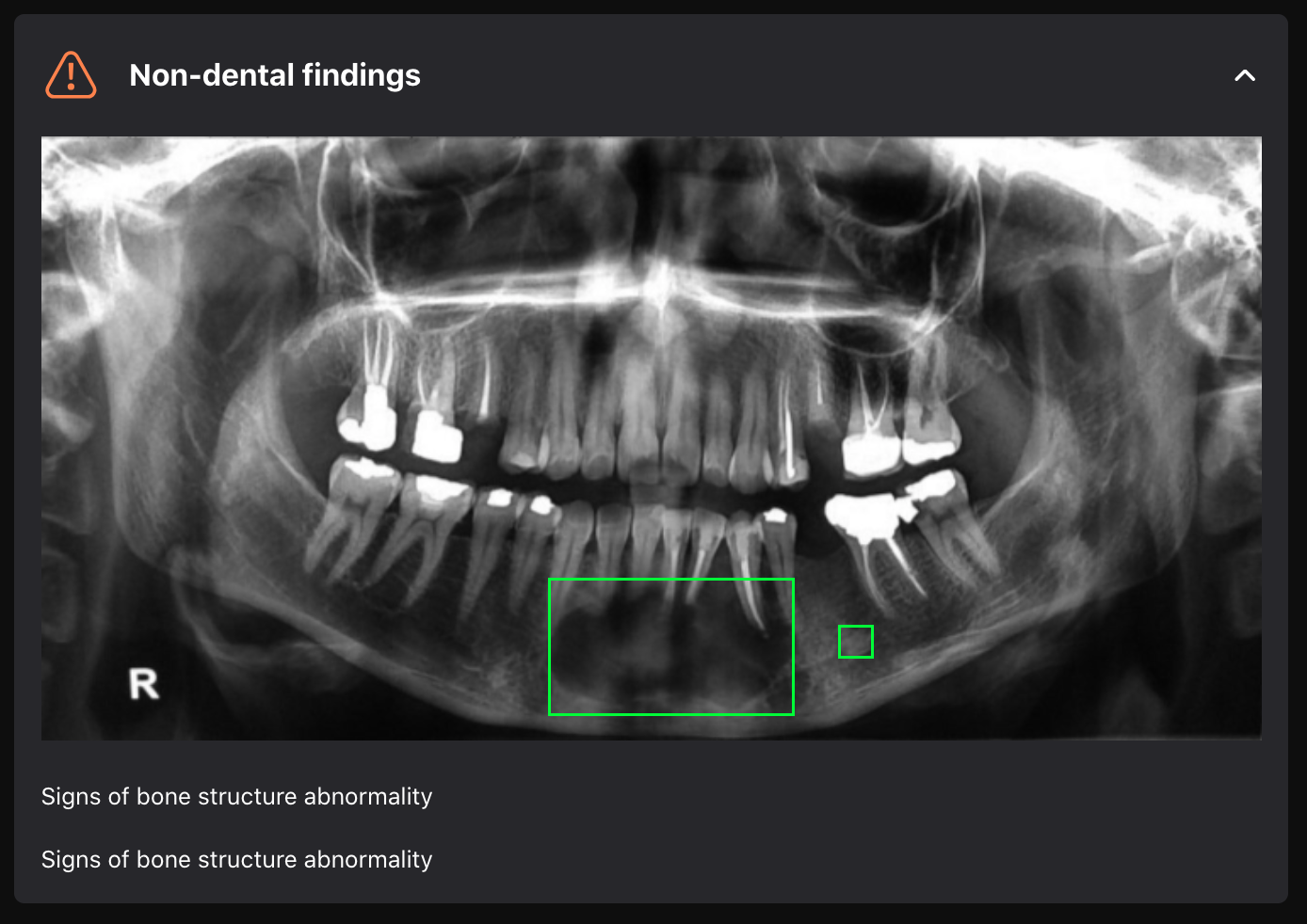
Complex analysis of the maxillofacial region
Diagnocat not only identifies dental conditions but also non-dental findings in the maxillofacial region, such as signs of abnormalities of the bone structure and maxillary sinus.
Printed report
The report is generated in PDF format and can be emailed or printed. Teeth requiring treatment are highlighted in red. A detailed radiological report complements the treatment plan and allows the patient to visually see the connection between the status of their oral cavity and the necessary treatment measures.
OPG pre-filled report
Visualization
Instant Panoramic visualization
Dental chart
Color-coded dental chart – highlighting healthy, previously treated, and unhealthy teeth
Findings
The list of possible findings includes 25+ conditions such as dental calculus, signs of caries, periapical lesion, periodontal bone loss, sinus and bone structure abnormalities, impactions, visible defects of previous endodontic treatment, lack of interproximal tooth contact among a variety of conditions aimed at enhancing the productivity of dental professionals
Smart view
Detailed single tooth smart view on the panoramic image
Recommendations
Referral recommendations
Printable reports
Printable reports for the medical record and improved communication with your patient
Explore more Diagnocat products
CBCT Segmentation

Cloud storage and Viewer

Collaboration Tool*

Specialists Reports*

Superimposition


